. 2005 Apr 5;102(14):5020-5.
doi: 10.1073/pnas.0501051102. Epub 2005 Mar 17.

S3b amino acid residues do not shuttle across the bilayer in voltage-dependent Shaker K+ channels

Affiliations

S3b amino acid residues do not shuttle across the bilayer in voltage-dependent Shaker K+ channels

Carlos Gonzalez et al. Proc Natl Acad Sci U S A. .

Abstract

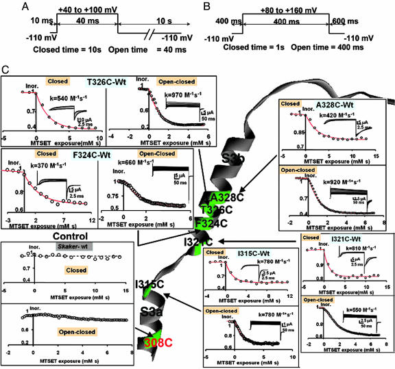
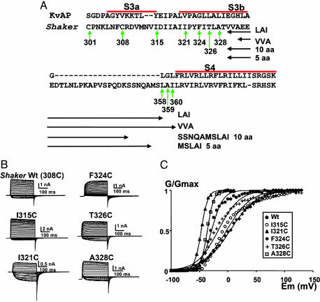
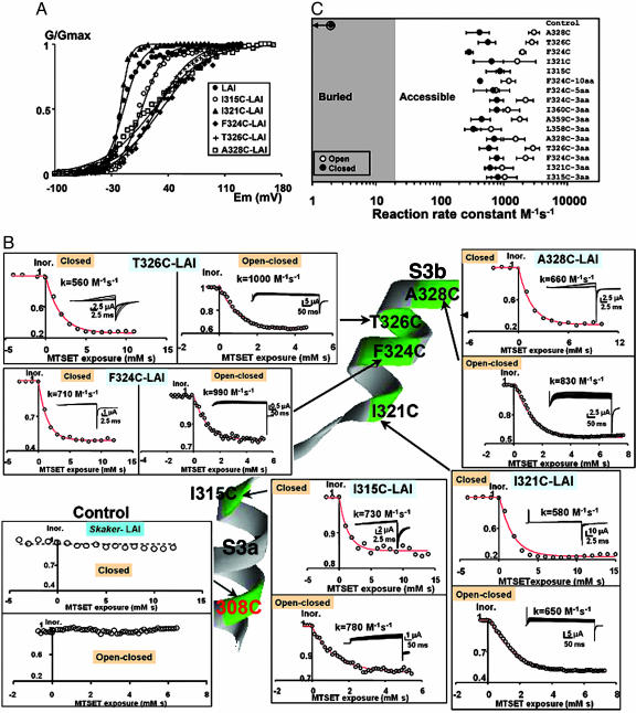
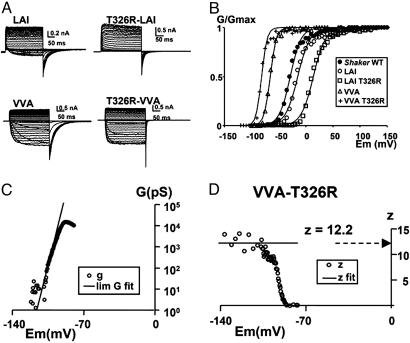
In voltage-dependent channels, positive charges contained within the S4 domain are the voltage-sensing elements. The "voltage-sensor paddle" gating mechanism proposed for the KvAP K+ channel has been the subject of intense discussion regarding its general applicability to the family of voltage-gated channels. In this model, the voltage sensor composed of the S3b and the S4 segment shuttles across the lipid bilayer during channel activation. Guided by this mechanism, we assessed here the accessibility of residues in the S3 segment of the Shaker K+ channel by using cysteine-scanning mutagenesis. Mutants expressed robust K+ currents in Xenopus oocytes and reacted with methanethiosulfonate ethyltrimethylammonium in both closed and open conformations of the channel. Because Shaker has a long S3-S4 linker segment, we generated a deletion mutant with only three residues to emulate the KvAP structure. In this short linker mutant, all of the tested residues in the S3b were accessible to methanethiosulfonate ethyltrimethylammonium in both closed and open conformations. Because the S3b moves together with the S4 domain in the paddle model, we tested the effects of deleting two negative charges or adding a positive charge to this region of the channel. We found that altering the S3b net charge does not modify the total gating charge involved in channel activation. We conclude that the S3b segment is always exposed to the external milieu of the Shaker K+ channel. Our results are incompatible with any model involving a large membrane displacement of segment S3b.


Figures

Fig. 2.
Fig. 2.
Voltage protocols for the assay of MTSET modification. (A) Closed protocol. The oocyte was clamped to a holding potential of –110 mV, channels were opened by applying a test pulse to either 40 or 100 mV with a duration of 40 ms, and the repetition rate was a pulse every 10 s. The magnitude of the test pulse was chosen by considering the characteristics of the voltage activation curve of the mutant channel. MTSET was added to the external side to a final concentration of 100 μM. Currents were measured by using the cut-open oocyte voltage-clamp technique. (B) Open/closed protocol. Channels were kept open by applying an 80–120 mV pulse with a 400-ms duration from a holding potential of –110. The total time in the closed state was 1 s. MTSET was added to the external side to a final concentration of 100 μM. (C) External accessibility of cysteines to MTSET. Cysteines were in positions 328, 326, 324, and 321 in the S3b segment and position 315 in the C terminus of segment S3a. All tested positions were accessible to MTSET in both the open/closed and the closed states. Rate constants expressed in M–1·s–1 are given in the figure for a representative experiment of each of the mutants. Note that the control Shaker channel contains a cysteine in position 308 that is inaccessible to the thiol reagent.
Fig. 1.
Fig. 1.
Shaker cysteine mutants express robust voltage-dependent currents. (A) Partial amino acid sequence of Shaker. Segments S3a, S3b, and S4 (bold) and the bar above the segments are assigned after Jiang et al. (26). Vertical green arrows show the native cysteines and the positions of the point mutations for cysteines. Horizontal double-headed arrows mark the four deleted regions. (B) Functional expression in Xenopus oocytes of cysteine mutants made in a ShakerΔ background. The holding potential was –100 mV, and the membrane was pulsed to voltages between –70 to +130 mV in 5 mV increments, followed by a step to –60 mV for ShakerΔ and the I315C, F324C, and T326C mutants. For the I321C and A328C, the holding potential was –100 mV and the membrane was pulsed to voltages between –100 to +10 mV in 5-mV increments, followed by a step to –100 mV. All current measurements were performed by using cell-attached patch–clamp technique. (C) Effects on the voltage activation induced by the mutation of S3 amino acid residues in a ShakerΔ background to cysteines. •, ShakerΔ WT; ○, I315C; ▴, I321C; ♦, F324C; +, T326C; □, A328C. Each point is the average of determinations on four to seven separate patches. Solid lines were drawn by using Eq. 1. V1/2 values were –29 ± 5 mV, –2 ± 16 mV, –47 ± 1 mV, 4 ± 15 mV, –17 ± 12 mV, and –36 ± 6 mV for ShakerΔ, I315C, I321C, F324C, T326C, and A328C, respectively. The equivalent numbers of gating charges, zeq, were 2.5 ± 1.2, 1.2 ± 0.9, 4.4 ± 0.1, 1.1 ± 0.2, 1.3 ± 0.6, and 4.5 ± 1.6 for ShakerΔ, I315C, I321C, F324C, T326C, and A328C, respectively.
Fig. 3.
Fig. 3.
Accessibility of S3b residues of short S3–S4 linker mutants. (A) Effects on the voltage-dependent activation in the S3 cysteine mutants with short-linker (LAI) background. •, LAI (control); ○, I315C; ▴, I321C; ♦, F324C; +, T326C; □, A328C. Each point is the average of determinations on four to seven separate patches. Solid lines were drawn by using Eq. 1. V1/2 values were –15 ± 8 mV, 7 ± 2 mV, –19 ± 2 mV, 32 ± 1 mV, 26 ± 5 mV, and 22 ± 9 mV for LAI (control), I315C, I321C, F324C, T326C, and A328C, respectively. The equivalent number of gating charges, zeq, was 2.2 ± 0.5, 1.8 ± 0.4, 4.5 ± 1.3, 0.9 ± 0.04, 1.1 ± 0.4, and 0.8 ± 0.4 for LAI (control), I315C, I321C, F324C, T326C, and A328C, respectively. (B) External accessibility of cysteines to MTSET in a short linker Shaker channel (LAI). Cysteines were in positions 328, 326, 324, and 321 in the S3b segment and in position 315 in C-terminal end of S3a. All tested positions were accessible to MTSET in both the open/closed and the closed states. Rates expressed in M–1·s–1 are given for representative experiments on each of the mutants. Note that the control LAI channel contains a cysteine in position 308 that is inaccessible to the thiol reagent. (C) State-dependent accessibility of the different cysteine mutants. Second-order rate constants are plotted for cysteine modification in the closed (•) and open (○) states. Mutant labeled F324-C10aa is a shortened linker Shaker mutant containing a S3–S4 linker of 10 aa (SSNQAMSLAI; see Fig. 1 A) and a cysteine in position 324. Mutant labeled F324C-5aa is a short linker Shaker mutant containing a S3–S4 linker of 5 aa (MSLAI; see Fig. 1 A) and a cysteine in position 324. LAC, LCI, and CAI are short linker Shaker mutants containing a S3–S4 linker of 3 aa and cysteines in the indicated positions. Each point is the mean of three or more determinations, and rates are expressed in M–1·s–1.
Fig. 4.
Fig. 4.
Mutation T326R does not increment gating charge. (A) Potassium current carried by T326R mutation in LAI and VVA S3–S4 linkers of mutant Shaker channels. Holding potentials were –100 and –130 mV for the LAI and VVA mutants, respectively. The membrane was pulsed to voltages between –70 to +30 mV in 5-mV increments, followed by a step to –60 mV for the LAI mutants and –120 to +80 mV in 5-mV increments and then by a step to –110 mV. All mutants were recorded in cell-attached patch–clamp configuration. (B) Relative conductance as a function of pulse potential of the mutants in A, measured from the tail currents. The solid lines were drawn by using Eq. 1. V1/2 were –29 ± 5 mV, –17 ± 9 mV, 13 ± 8 mV, –68 ± 14 mV, and –83 ± 16 mV for ShakerΔ, LAI, T326R-LAI, VVA, and T326R-VVA, respectively. Limiting-slope analysis in charged S3b short linker mutant channels. (C) Semilogarithmic plot of the G vs. V relationship of the VVA-T326R mutant. The solid line indicates the fitting of the low-probability data to determine the limiting value of zeff. (D) z vs. V plot. The circles correspond to the values of z calculated from Eq. 3. The solid line indicates the value of zeff obtained from the monoexponential fit of conductance vs. voltage relationship for conductance values <0.001 (Eq. 2). Average values of zeff for the VVA mutant was 11.5 ± 0.5 (5) and form the VVA-T326R mutant was 12.5 ± 0.5 (5).

Comment in

  • How ion channels sense membrane potential.
    Horn R. Horn R. Proc Natl Acad Sci U S A. 2005 Apr 5;102(14):4929-30. doi: 10.1073/pnas.0501640102. Epub 2005 Mar 28. Proc Natl Acad Sci U S A. 2005. PMID: 15795366 Free PMC article. No abstract available.

Similar articles

Cited by

References

    1. Aggarwal, S. K. & MacKinnon, R. (1996) Neuron 16, 1169–1177. - PubMed
    1. Seoh, S. A., Sigg, D., Papazian, D. M. & Bezanilla, F. (1996) Neuron 16, 1159–1167. - PubMed
    1. Starace, D. M. & Bezanilla, F. (2001) J. Gen. Physiol. 117, 469–490. - PMC - PubMed
    1. Starace, D. M. & Bezanilla, F. (2004) Nature 427, 548–553. - PubMed
    1. Swartz, K. J. (2004) Nat. Rev. Neurosci. 5, 905–916. - PubMed

Publication types

MeSH terms

LinkOut - more resources