

Cardioprotective effects of peroxisome proliferator activated receptor gamma activators on acute myocarditis: anti-inflammatory actions associated with nuclear factor kappaB blockade
- PMID: 15774612
- PMCID: PMC1769084
- DOI: 10.1136/hrt.2004.046292
Cardioprotective effects of peroxisome proliferator activated receptor gamma activators on acute myocarditis: anti-inflammatory actions associated with nuclear factor kappaB blockade
Abstract
Objective: To test the hypothesis that activation of peroxisome proliferator activated receptor gamma (PPAR-gamma) reduces experimental autoimmune myocarditis (EAM) associated with inhibitor kappaB (IkappaB) alpha induction, blockade of nuclear factor kappaB (NF-kappaB), and inhibition of inflammatory cytokine expression.
Methods: EAM was induced in Lewis rats by immunisation with porcine cardiac myosin. PPAR-gamma activators 15-deoxy-Delta(12,14)-prostaglandin J2 (15d-PGJ2) and pioglitazone (PIO) were administered to rats with EAM.
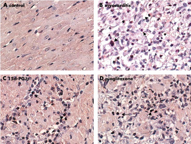
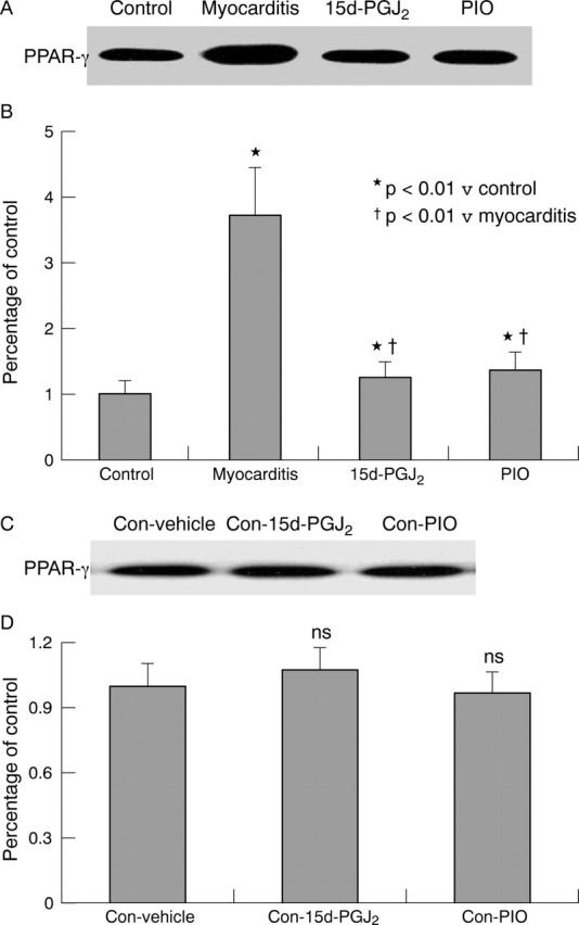
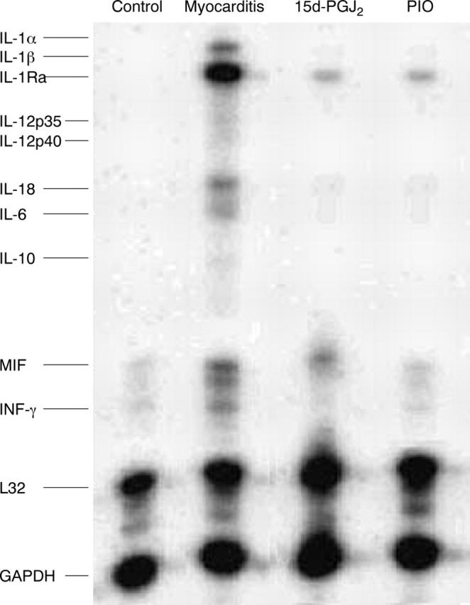
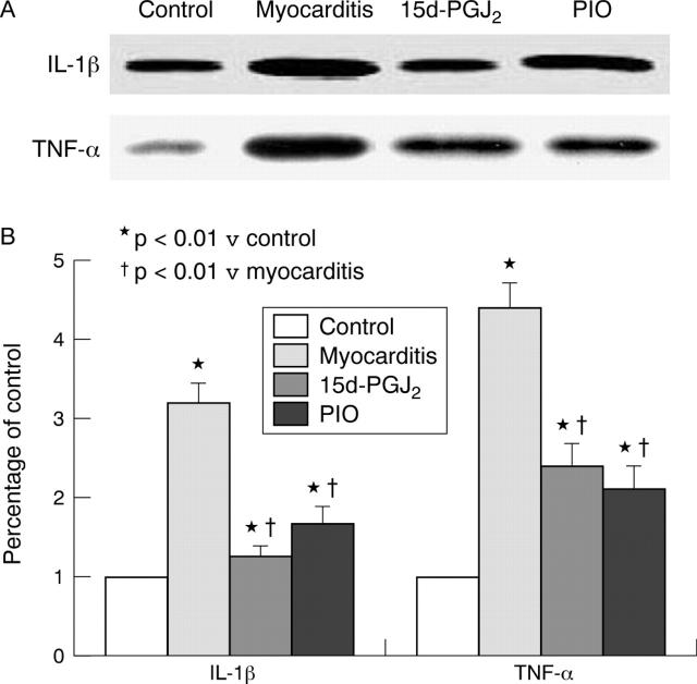
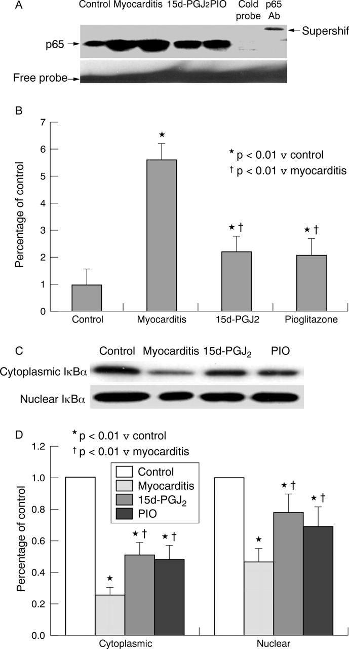
Results: Enhanced PPAR-gamma expression was prominently stained in the nuclear and perinuclear regions of infiltrating inflammatory cells. Administration of 15d-PGJ2 and PIO greatly reduced the severity of myocarditis and suppressed myocardial mRNA and protein expression of inflammatory cytokines in rats with EAM. In addition, treatment with PPAR-gamma activators enhanced IkappaB concentrations in the cytoplasmic fractions and nuclear fractions from inflammatory myocardium. Concurrently, NF-kappaB was greatly activated in myocarditis; this activation was blocked in the 15d-PGJ2 treated and PIO treated groups.
Conclusions: PPAR-gamma may have a role in the pathophysiology of EAM. Because an increase in IkappaB expression and inhibition of translocation of the NF-kappaB subunit p65 to the nucleus in inflammatory cells correlated with the protective effects of PPAR-gamma activators, these results suggest that PPAR-gamma activators act sequentially through PPAR-gamma activation, IkappaB induction, blockade of NF-kappaB activation, and inhibition of inflammatory cytokine expression. These results suggest that PPAR-gamma activators such as 15d-PGJ2 and PIO may have the potential to modulate human inflammatory heart diseases such as myocarditis.
Figures
Similar articles
-
Peroxisome proliferation-activated receptor-gamma ligands ameliorate experimental autoimmune myocarditis.Cardiovasc Res. 2003 Sep 1;59(3):685-94. doi: 10.1016/s0008-6363(03)00457-7. Cardiovasc Res. 2003. PMID: 14499870
-
[Peroxisome proliferation-activated receptor-gamma ligands ameliorate autoimmune myocarditis associated with inhibition of T cell immunity].Zhonghua Yi Xue Za Zhi. 2003 Dec 10;83(23):2067-72. Zhonghua Yi Xue Za Zhi. 2003. PMID: 14703419 Chinese.
-
Peroxisome proliferator-activated receptor-gamma ligands ameliorate experimental autoimmune myocarditis associated with inhibition of self-sensitive T cells.J Cardiovasc Pharmacol. 2004 Jun;43(6):868-75. doi: 10.1097/00005344-200406000-00017. J Cardiovasc Pharmacol. 2004. PMID: 15167281
-
15-Deoxy-Δ-12,14-prostaglandin J2 effects in vascular smooth muscle cells: Implications in vascular smooth muscle cell proliferation and contractility.Prostaglandins Other Lipid Mediat. 2021 Oct;156:106583. doi: 10.1016/j.prostaglandins.2021.106583. Epub 2021 Jul 29. Prostaglandins Other Lipid Mediat. 2021. PMID: 34332056 Review.
-
Understanding the peroxisome proliferator-activated receptor gamma (PPAR-γ) role in periodontitis and diabetes mellitus: A molecular perspective.Biochem Pharmacol. 2025 Jul;237:116908. doi: 10.1016/j.bcp.2025.116908. Epub 2025 Mar 27. Biochem Pharmacol. 2025. PMID: 40157459 Review.
Cited by
-
Enhanced effects of cigarette smoke extract on inflammatory cytokine expression in IL-1β-activated human mast cells were inhibited by Baicalein via regulation of the NF-κB pathway.Clin Mol Allergy. 2012 Feb 6;10:3. doi: 10.1186/1476-7961-10-3. Clin Mol Allergy. 2012. PMID: 22309647 Free PMC article.
-
Simvastatin activates the PPARγ-dependent pathway to prevent left ventricular hypertrophy associated with inhibition of RhoA signaling.Tex Heart Inst J. 2013;40(2):140-7. Tex Heart Inst J. 2013. PMID: 23678211 Free PMC article.
-
Baicalein inhibits IL-1beta- and TNF-alpha-induced inflammatory cytokine production from human mast cells via regulation of the NF-kappaB pathway.Clin Mol Allergy. 2007 Nov 26;5:5. doi: 10.1186/1476-7961-5-5. Clin Mol Allergy. 2007. PMID: 18039391 Free PMC article.
-
Multiple Interactions between Peroxisome Proliferators-Activated Receptors and the Ubiquitin-Proteasome System and Implications for Cancer Pathogenesis.PPAR Res. 2008;2008:195065. doi: 10.1155/2008/195065. PPAR Res. 2008. PMID: 18551186 Free PMC article.
-
Targeting PPARs Signaling Pathways in Cardiotoxicity by Natural Compounds.Cardiovasc Toxicol. 2022 Apr;22(4):281-291. doi: 10.1007/s12012-021-09715-5. Epub 2022 Jan 24. Cardiovasc Toxicol. 2022. PMID: 35067839 Review.
References
-
- Neumann FJ, Ott I, Gawaz M, et al. Cardiac release of cytokines and inflammatory responses in acute myocardial infarction. Circulation 1995;92:748–55. - PubMed
-
- Yuan Z, Shioji K, Kihara Y, et al. Cardioprotective effects of carvedilol on acute autoimmune myocarditis: anti-inflammatory effects associated with antioxidant property. Am J Physiol Heart Circ Physiol 2004;286:83–90. - PubMed
Publication types
MeSH terms
Substances
LinkOut - more resources
Full Text Sources
Other Literature Sources
Medical
Molecular Biology Databases