. 2005 Apr 1;19(7):815-26.
doi: 10.1101/gad.1284005. Epub 2005 Mar 17.

Histone methyltransferases G9a and GLP form heteromeric complexes and are both crucial for methylation of euchromatin at H3-K9

Affiliations

Histone methyltransferases G9a and GLP form heteromeric complexes and are both crucial for methylation of euchromatin at H3-K9

Makoto Tachibana et al. Genes Dev. .

Abstract

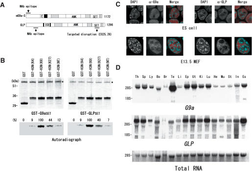
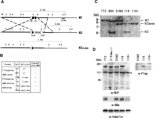
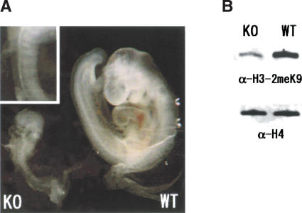
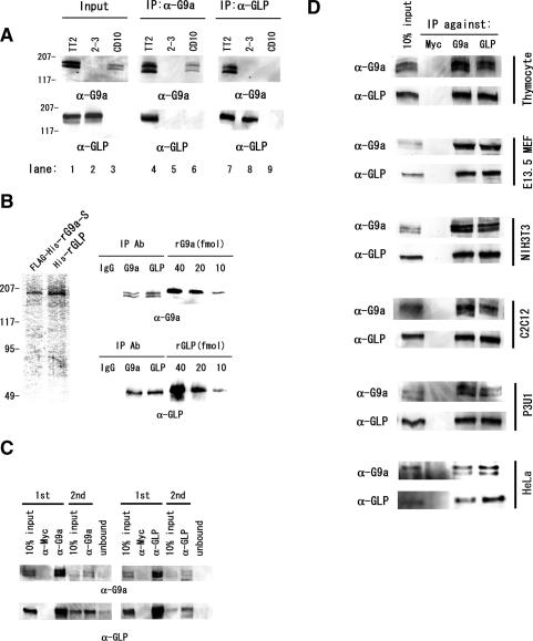
Histone H3 Lys 9 (H3-K9) methylation is a crucial epigenetic mark for transcriptional silencing. G9a is the major mammalian H3-K9 methyltransferase that targets euchromatic regions and is essential for murine embryogenesis. There is a single G9a-related methyltransferase in mammals, called GLP/Eu-HMTase1. Here we show that GLP is also important for H3-K9 methylation of mouse euchromatin. GLP-deficiency led to embryonic lethality, a severe reduction of H3-K9 mono- and dimethylation, the induction of Mage-a gene expression, and HP1 relocalization in embryonic stem cells, all of which were phenotypes of G9a-deficiency. Furthermore, we show that G9a and GLP formed a stoichiometric heteromeric complex in a wide variety of cell types. Biochemical analyses revealed that formation of the G9a/GLP complex was dependent on their enzymatic SET domains. Taken together, our new findings revealed that G9a and GLP cooperatively exert H3-K9 methyltransferase function in vivo, likely through the formation of higher-order heteromeric complexes.


Figures

Figure 1.
Figure 1.
Molecular and expression characteristics of G9a and GLP. (A) Molecular structures of mouse G9a-S and GLP. Black boxes represent putative nuclear localization sequences. Dark boxes represent cysteine-rich regions including pre- and post-SET domains. (ANK) Ankyrin repeats; (SET) SET domains; (E) poly glutamic acid stretch; (ED) acidic region composed of glutamic and aspartic acid repeats. (B) The SET domains of G9a and GLP share the same substrate specificity. HMTase activities of the SET domains of G9a (amino acids 878–1172 for G9a-S, left) and GLP (amino acids 1002–1296, right). The GST-SET domains were incubated with 14C-labeled SAM and several GST-fused H3-N-terminal proteins (1–57) as substrates. Mutant substrates carry double K-to-R substitutions as follows: 4K, (9 + 27)R; K9, K(4 + 27)R; K27, K(4 + 9)R. (NT) Triple K-to-R substitutions. Asterisks indicate GST-SET domain proteins. (C) Nuclear distributions of G9a and GLP. Wild-type ES cells (top) and mouse primary fibroblasts from wild-type E13.5 embryos (bottom) were stained with DAPI (right, blue) and specific antibodies against G9a or GLP (red). (D) Expression profiles of G9a and GLP transcripts in mouse tissues. Four micrograms of total RNA were probed with radio-labeled G9a (top) and GLP cDNA (middle). (Bottom) 28S ribosomal RNA was visualized by ethidium bromide staining as a loading control. (Th) Thymus; (Sp) spleen; (Ly) lymph node; (Bo) bone marrow; (Br) brain; (Te) testis; (Li) liver; (Ep) epididymis; (Ut) uterus; (Ki) kidney; (Lu) lung; (He) heart; (Mu) skeletal muscle; (St) stomach; (In) intestine; (Es) ES cells (TT2).
Figure 2.
Figure 2.
Generation of GLP-deficient mice and ES cells. (A) Partial restriction maps of the mouse GLP locus (exons 25 and 26, top), and the targeted GLP locus containing a neomycin cassette (middle) are shown. (Bottom) The neomycin cassette can be excised from the targeted locus for disruption of the second allele. The portions of the endogenous locus that were used for constructing the targeting plasmid are shown as thick lines. (Xh) XhoI; (H) HindIII; (X) XbaI; (A) ArtII; (B) BamHI. (B) Flow diagram for the establishment of GLP-deficient ES cells. To disrupt GLP conditionally with Cre-recombinase, loxP-flanked (flox) Flag-GLP-cDNA was introduced before the second GLP targeting. The 118 cells, which expressed only exogenous Flag-GLP-cDNA, were treated with 5′OHT to excise the Flag-GLP-cDNA. The resultant cells (118+) successfully carried a GLP-null genotype. (C) Southern blot analyses of GLP alleles described in A. Disruption of the GLP locus was confirmed by Southern blot analysis using BamHI/HindIII-digested DNA probed with a genomic portion located outside of the targeting construct as shown in A.(D) GLP deficiency was confirmed by Western blot analyses using total lysates corresponding to 105 cells. Endogenous GLP protein was detected at a molecular weight of ∼170 kDa. (Top) Asterisks represent nonspecific signals detected by the secondary antibodies. (Bottom) Tubulin contents were also determined as a loading control.
Figure 3.
Figure 3.
GLP-deficiency results in embryonic-lethality. (A) The gross morphology of a typical GLP-deficient embryo (left) and a wild-type sibling (right) at E9.5. GLP-deficient embryos were severely growth-retarded, containing only 4–6 somites (enlarged in upper left), while wild-type siblings had 21–25 somites. (B) Acid-extracted histones from E9.5 whole embryos were separated by SDS-PAGE and followed by immunoblotting with anti-dimethyl H3-K9 (top) or anti-H4 antibodies (bottom). The levels of H3-K9 dimethylation were drastically reduced in GLP-deficient whole embryos.
Figure 4.
Figure 4.
Phenotypes of GLP-deficient ES cells. (A,B) The H3-K9 methylation status of TT2 (wild type), G9a-deficient (2-3), and GLP-deficient cells (CD10) was analyzed by Western blot (A) and immunostaining analyses (B). (A, left) GLP protein was absent from in CD10 cells. The reduction in levels of H3-K9 mono-and dimethylation in GLP-deficient cells were indistinguishable from that observed in 2-3 cells. Overall H3-K9 trimethylation at pericentric heterochromatin was unaffected in the mutant cells. (C) The G9a or GLP mutations alter the nuclear distribution of HP1 proteins. Mutant cell nuclei were stained with specific antibodies against three HP1 isoforms (α, left; β, middle; γ, right panels). In G9a- and GLP-mutant cells, euchromatic staining profiles of HP1 had significantly disappeared, but were enriched at pericentic loci. (D) GLP suppressed Mage-a gene expression. (Left) Five micrograms of total RNA were separated and probed with radiolabeled Mage-a cDNA. Fixed and sonicated chromatin of TT2, 2-3, and CD10 ES cells were immunoprecipitated with the indicated Abs and applied to the semiquantitative assay. (Right) H3-K9 dimethylation was reduced and H3-K4 dimethylation was increased on the Mage-a2 promoter region in G9a- and GLP-deficient cells. (E) GLP-cDNA introduction can rescue GLP-deficient phenotypes. A GLP-cDNA driven by the chicken β actin promoter was introduced stably into GLP-deficient cells. (Left panels) In rescued clones L2 and L6, which expressed GLP at levels comparable to those observed in wild-type cells, the protein stability of G9a was recovered. Similarly, dimethyl H3-K9 levels and Mage-a gene suppression were restored (middle and right panels, respectively).
Figure 5.
Figure 5.
G9a and GLP form heteromeric complexes. (A) Endogenous G9a and GLP were coprecipitated with specific antibodies against G9a or GLP. (Middle panels) Only the anti-G9a immunocomplexes from wild-type nuclear extracts contain GLP. (Right panels) Similarly, immune complexes from wild-type cells obtained with anti-GLP antibody also contain G9a. (B) Quantitative analyses of anti-G9a and anti-GLP immunocomplexes. (Left) Semipurified recombinant G9a and GLP from virus-infected insect cells were used as protein standards. Amounts of G9a or GLP protein content in immunocomplexes from wild-type nuclear extracts were measured by immunoblotting with precalibrated recombinant G9a or GLP. (Right) Each of the anti-G9a or anti-GLP immunocomplex contains between 10 and 20 fmol of each molecule. (C) G9a/GLP predominantly form heteromeric complexes. Two sequential immunodepletions using anti-G9a (left) or anti-GLP (right) led to a drastic reduction of both proteins from nuclear extracts. (D) G9a and GLP ubiquitously form a heteromeric complex. Anti-G9a and anti-GLP immunocomplexes were prepared from mouse adult thymocytes, E13.5 primary fibroblasts, the NIH3T3 cell line, the C2C12 myocyte cell line, the myeloma cell line P3U1, and the human cell line HeLa and were subjected to Western blot analysis. G9a and GLP formed a heteromeric complex in all murine and human cells tested and this heteromeric complexes were the predominant form.
Figure 6.
Figure 6.
G9a/GLP heteromeric complexes require their SET domains. (A,B) Specificity and interaction domain analysis for the G9a and GLP proteins. Flag-G9a-S (A) and GLP (B) were coexpressed with several other EGFP-tagged, H3-K9 HMTases specified at the top of the figure. Immunoprecipitates were collected with control anti-Myc antibody or anti-Flag antibody. Neither G9a nor GLP interacted with Suv39h1 or ESET. Deletion analysis indicated that the interaction between G9a and GLP required their SET domains. (C) Interaction of recombinant G9a and GLP in insect cells. Vaculo viruses encoding G9a or GLP were coinfected (left) or independently infected (right) to Sf9 cells, and recombinant G9a and GLP were collected with anti-G9a or anti-GLP antibodies. G9a and GLP could interact in insect cells when the corresponding viruses were coinfected (left panels), whereas the mixture of lysate from independently infected cells could not produce a G9a/GLP heterotypic interaction. (D) G9a and GLP can interact homotypically. Flag-G9a-S (left) or Flag-GLP (right) were coexpressed with their EGFP-tagged versions, and collected with control anti-Myc antibody or anti-Flag antibody. Homomeric interaction of G9a or GLP was also dependent on their SET domains. (E,F) SET domains are necessary and sufficient for dimerization. (E) Generation of epitope-tagged SET domains. G9a-SET (amino acids 878–1172 for G9a-S) and that of GLP (amino acids 1002–1296) carrying Myc- or Flag-tag were expressed in HEK 293T cells and detcted with anti-tag antibodies. (F) Interaction between the SET domains. Combinations of SET domain proteins indicated at top were coexpressed in HEK 293T cells and subjected to immunocomplex analyses using control IgG1 or anti-Flag antibody. (Top) Immunoblot analysis with anti-Myc indicated that the SET domains can hetero- or homodimerize. For negative control, Myc-G9a-SET and Flag-EGFP proteins were coexpressed and subjected to immunocomplex analyses. (Bottom) Neither anti-Myc nor anti-Flag immunoprecipitates contained partner molecule. Asterisk indicates protein-G signals extracted from resin.

References

    1. Aagaard L., Laible, G., Selenko, P., Schmid, M., Dorn, R., Schotta, G., Kuhfittig, S., Wolf, A., Lebersorger, A., Singh, P.B., et al. 1999. Functional mammalian homologs of the Drosophila PEV-modifier Su(var)3-9 encode centromere-associated proteins which complex with the heterochromatin component M31. EMBO J. 18: 1923–1938. - PMC - PubMed
    1. Bannister A.J., Zegerman, P., Partridge, J.F., Miska, E.A., Thomas, J.O., Allshire, R.C., and Kouzarides, T. 2001. Selective recognition of methylated lysine 9 on histone H3 by the HP1 chromo domain. Nature 410: 120–124. - PubMed
    1. Cao R. and Zhang, Y. 2004a. The functions of E(Z)/EZH2-mediated methylation of lysine 27 in histone H3. Curr. Opin. Genet. Dev. 14: 155–164. - PubMed
    1. ____. 2004b. SUZ12 is required for both the histone methyltransferase activity and the silencing function of the EED–EZH2 complex. Mol. Cell 15: 57–67. - PubMed
    1. Cheutin T., McNairn, A.J., Jenuwein, T., Gilbert, D.M., Singh, P.B., and Misteli, T. 2003. Maintenance of stable heterochromatin domains by dynamic HP1 binding. Science 299: 721–725. - PubMed

Publication types