. 2005 Apr;187(7):2377-85.
doi: 10.1128/JB.187.7.2377-2385.2005.

The gusBC genes of Escherichia coli encode a glucuronide transport system

Affiliations

The gusBC genes of Escherichia coli encode a glucuronide transport system

Wei-Jun Liang et al. J Bacteriol. 2005 Apr.

Abstract

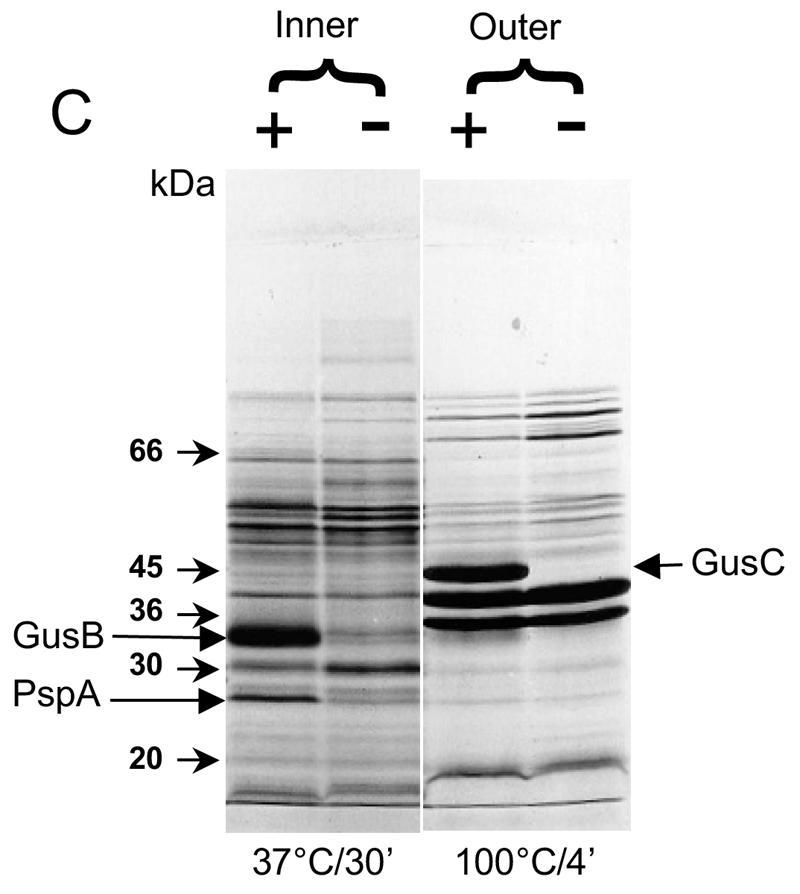
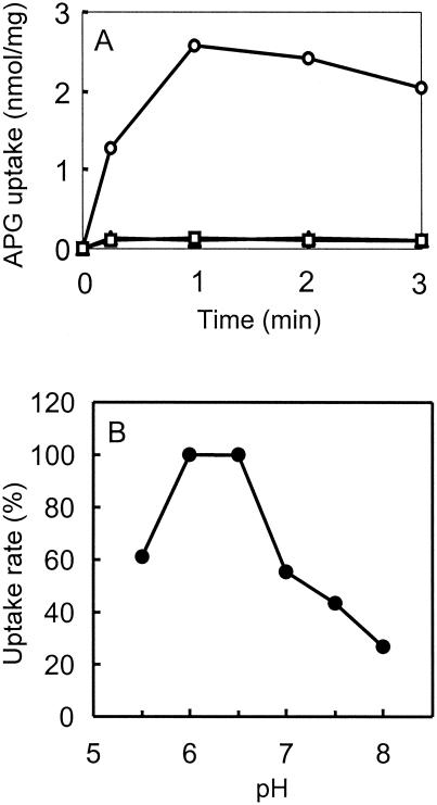
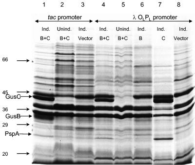
Two genes, gusB and gusC, from a natural fecal isolate of Escherichia coli are shown to encode proteins responsible for transport of beta-glucuronides with synthetic [(14)C]phenyl-1-thio-beta-d-glucuronide as the substrate. These genes are located in the gus operon downstream of the gusA gene on the E. coli genome, and their expression is induced by a variety of beta-d-glucuronides. Measurements of transport in right-side-out subcellular vesicles show the system has the characteristics of secondary active transport energized by the respiration-generated proton motive force. When the genes were cloned together downstream of the tac operator-promoter in the plasmid pTTQ18 expression vector, transport activity was increased considerably with isopropylthiogalactopyranoside as the inducer. Amplified expression of the GusB and GusC proteins enabled visualization and identification by N-terminal sequencing of both proteins, which migrated at ca. 32 kDa and 44 kDa, respectively. Separate expression of the GusB protein showed that it is essential for glucuronide transport and is located in the inner membrane, while the GusC protein does not catalyze transport but assists in an as yet unknown manner and is located in the outer membrane. The output of glucuronides as waste by mammals and uptake for nutrition by gut bacteria or reabsorption by the mammalian host is discussed.


Figures

FIG. 1.
FIG. 1.
Cloning of the gus operon. A 7,740-bp PstI-HindIII fragment containing the gus operon from a natural E. coli fecal isolate (CE1) was cloned into the PstI and HindIII restriction sites (not shown) of low-copy-number plasmid pLAFR3, generating plasmid pKW219. The corresponding fragment of the gus operon from E. coli K-12 was also cloned to generate plasmid pKW220 (not shown). A BstYI fragment (3,368 bp) containing the gusBCCE1 genes was subcloned into plasmid pAD284 at the HpaI site, generating plasmid pWJL4, so that the gene expression is under the control of the λ OLPL promoter. Plasmids containing the gusBCE1 gene (pWJL6) and the gusCCE1 gene (pWJL11) were generated from plasmid pWJL4 by a combination of restriction digestions (see Materials and Methods for details). The base pair numbers are those listed in GenBank (accession number M14641), while the base pair numbers in brackets correspond to those listed in the genome sequence of E. coli MG1655 (3).
FIG. 2.
FIG. 2.
[14C]-Phenyl-thio-β-d-glucuronide (PTG) transport in different host-vector systems. Each line represents curve fits of mean values of triplicate analyses at each of the time points; error bars are standard deviations. PTG was used at 50 μM. (A) Induction of the complete gusCE1 operon on plasmid pKW219 or the gusK-12 operon on plasmid pKW220. The host E. coli strain KW1 has the entire gus operon deleted. Cells were induced with 1 mM p-nitrophenyl-β-d-glucuronide for 40 min during logarithmic growth. Solid upward triangles, PTG uptake into cells with induced gusABCCE1 operon (line represents curve fits); open downward triangles, cells with uninduced gusABCCE1 operon; solid squares, cells with induced gusK-12 operon; solid circles, vector control [KW1(pLARF3)]; solid downward triangle, strain control (KW1). (B) Expression of the gusBCE1 and gusCCE1 genes under control of the λ OLPL promoter in the λ lysogen strain AR120. Strains were induced with 40 μg of nalidixic acid per ml for 3 h at mid-log phase. Solid squares, cells with induced gusBCCE1 genes on plasmid pWJL4; solid downward triangles, cells with uninduced gusBCCE1 genes on plasmid pWJL4; open squares, cells with induced gusBCE1 gene alone on plasmid pWJL6; open diamonds, cells with induced gusCCE1 gene alone on plasmid pWJL11; solid upward triangles, vector control [AR120(pAD284)]. (C) Expression of the gusBCE1 and gusCCE1 genes under control of the tac promoter. Strains of E. coli NO2947 containing the plasmids below were induced with 1 mM IPTG for 14 h after inoculation in minimal medium. Solid upward triangles, cells with induced gusBCCE1 genes on plasmid pWJL24; solid downward triangles, cells with uninduced gusBCCE1 genes on plasmid pWJL24; open upward triangles, cells with induced gusBCE1 gene alone on plasmid pWJL26; open diamonds, cells with induced gusCCE1 gene alone on plasmid pWJL31; solid circles, vector control [NO2947(pTTQ18)].
FIG. 3.
FIG. 3.
Appearance of putative GusB and GusC proteins in membrane preparations. (A) Total membrane proteins prepared from E. coli strain NO2947 harboring plasmids as indicated below were incubated in solubilization buffer at 37°C for 30 min; 45 μg of proteins was loaded onto each track of the SDS-12% bisacrylamide gel. Lane 1, protein size markers (in kilodaltons); lane 2, plasmid pWJL24 tac-gusBC induced; lane 3, pWJL24 tac-gusBC uninduced; lane 4, pTTQ18 induced control; lane 5, no plasmid in host cell control. (B) After solubilization conditions as shown, total membrane protein (45 μg) was loaded onto each track of the SDS-12% bisacrylamide gel. Lane B+C, pWJL24 tac-gusBCCE1 induced; lane B, pWJL26 tac-gusB CE1 induced; lane C, pWJL31 tac-gusCCE1 induced. Proteins in lanes 4, 5, and 6 were as same as those in lane 1, 2, and 3, respectively. (C) GusB occurs in the inner membrane and GusC in the outer membrane. Membrane fractionation was carried out by sucrose gradient and ultracentrifugationafter cell lysis by French press (20,000 lb/in2). Inner, inner membrane proteins solubilized at 37°C for 30 min; Outer, outer membrane proteins solubilized at 100°C for 4 min. +, proteins from NO2947(pWJL24) with induction of = tac-gusBCCE1; −, proteins from NO2947(pWJL24) without induction of tac-gusBCCE1.
FIG. 3.
FIG. 3.
Appearance of putative GusB and GusC proteins in membrane preparations. (A) Total membrane proteins prepared from E. coli strain NO2947 harboring plasmids as indicated below were incubated in solubilization buffer at 37°C for 30 min; 45 μg of proteins was loaded onto each track of the SDS-12% bisacrylamide gel. Lane 1, protein size markers (in kilodaltons); lane 2, plasmid pWJL24 tac-gusBC induced; lane 3, pWJL24 tac-gusBC uninduced; lane 4, pTTQ18 induced control; lane 5, no plasmid in host cell control. (B) After solubilization conditions as shown, total membrane protein (45 μg) was loaded onto each track of the SDS-12% bisacrylamide gel. Lane B+C, pWJL24 tac-gusBCCE1 induced; lane B, pWJL26 tac-gusB CE1 induced; lane C, pWJL31 tac-gusCCE1 induced. Proteins in lanes 4, 5, and 6 were as same as those in lane 1, 2, and 3, respectively. (C) GusB occurs in the inner membrane and GusC in the outer membrane. Membrane fractionation was carried out by sucrose gradient and ultracentrifugationafter cell lysis by French press (20,000 lb/in2). Inner, inner membrane proteins solubilized at 37°C for 30 min; Outer, outer membrane proteins solubilized at 100°C for 4 min. +, proteins from NO2947(pWJL24) with induction of = tac-gusBCCE1; −, proteins from NO2947(pWJL24) without induction of tac-gusBCCE1.
FIG. 4.
FIG. 4.
Energization of transport of glucuronides into right-side-out (RSO) subcellular vesicles. (A) Subcellular vesicles were prepared from E. coli NO2947(pWJL24) grown on minimal medium and induced with IPTG (see text). After incubation for 3 min with additions as below, 50 μM [14C]APG was added and transport was measured. Triangles, no addition; squares, plus 20 mM glycerol; circles, plus 20 mM ascorbate and 0.1 mM phenazine methosulfate. Similar results were obtained when 50 μM [14C]PTG was the substrate (not shown). The pH was 6.6. Points are means of duplicate measurement. (B) Samples of right-side-out vesicles were resuspended in 40 mM potassium phosphate buffer at the indicated pH. After 3 min of incubation, the initial rate of transport of 50 μM [14C]APG was measured with 20 mM ascorbate and 0.1 mM phenazine methosulfate for the respiratory substrate. Points are means of duplicates (15-s time point) expressed as a percentage of the highest value of 1.87 nmol/mg.
FIG. 5.
FIG. 5.
Expression of the GusB and GusC proteins in tac and OLPL promoter systems. Cells containing the plasmids indicated below were grown and membrane preparations were made; 1.0 mM IPTG was included for induction of the tacpromoter and 40 μg of nalidixic acid per ml for induction of the OLPL promoter where indicated. Total membrane protein was solubilized at 100°C for 4 min and 45 μg was loaded onto each track of the SDS--12% bisacrylamide gel. Lane 1, tac-gusBCCE1 induced; lane 2, tac-gusBCCE1 uninduced; lane 3, vector pTTQ18 plus IPTG; lane 4, OLPL gusBCCE1 induced; lane 5, OLPL gusBCCE1 uninduced; lane 6, OLPL gusBCE1 induced; lane 8, OLPL gusCCE1 induced; lane 10, vector pAD284 plus nalidixic acid.

Similar articles

Cited by

References

    1. Ahmed, S., and I. R. Booth. 1981. Quantitative measurements of the proton-motive force and its relation to steady state lactose accumulation in Escherichia coli. Biochem. J. 200:573-581. - PMC - PubMed
    1. Banhegyi, G., L. Braun, P. Marcolongo, M. Csala, R. Fulcer, J. Mandl, and A. Benedetti. 1996. Evidence for an UDP-glucuronic acid/phenol glucuronide antiport in rat liver microsomal vesicles. Biochem. J. 315:171-176. - PMC - PubMed
    1. Blattner, F. R., G. Plunkett, C. A. Bloch, N. T. Perna, V. Burland, M. Riley, J. Collado-Vides, J. D. Glasner, C. K. Rode, G. F. Mayhew, J. Gregor, N. W. Davis, H. A. Kirkpatrick, M. A. Goeden, D. J. Rose, B. Mau, and Y. Shao. 1997. The complete genome sequence of Escherichia coli K-12. Science 277:1453-1474. - PubMed
    1. Boos, W., and J. M. Lucht. 1996. Periplasmic binding protein-dependent ABC transporters, p. 1175-1209. In F. C. Neidhardt et al. (ed.) Escherichia coli and Salmonella: cellular and molecular biology, 2nd ed. ASM Press, Washington, D.C.
    1. Brissette, J. L., L. Weiner, T. L. Ripmaster, and P. Model. 1991. Characterization and sequence of the Escherichia coli stress-induced psp operon. J. Mol. Biol. 220:35-48. - PubMed

Publication types

MeSH terms

LinkOut - more resources