

Macular function and morphology after peeling of idiopathic epiretinal membrane with and without the assistance of indocyanine green
- PMID: 15774920
- PMCID: PMC1772583
- DOI: 10.1136/bjo.2004.051250
Macular function and morphology after peeling of idiopathic epiretinal membrane with and without the assistance of indocyanine green
Abstract
Aim: To investigate macular function and morphology after surgical removal of idiopathic epiretinal membrane (IEM) with and without assistance of indocyanine green (ICG).
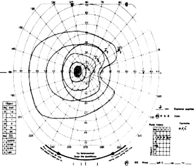
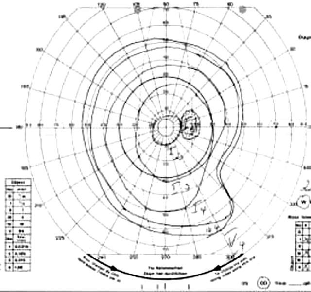
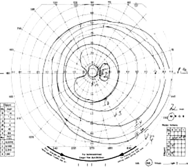
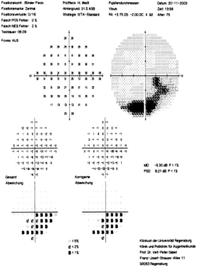
Methods: A retrospective study as a consecutive case series, of 39 patients with IEM. 39 patients, 23 female, 16 male, mean age 67 years, underwent standard three port pars plana vitrectomy with removal of epiretinal membrane. Two groups of patients were consecutively operated: in 20 patients ICG 0.1% in glucose 5% was used to stain the epiretinal membrane. 19 patients underwent the identical procedure but without use of ICG. Postoperative follow up was 1-92 months (mean 15.5 months). Functional outcome was assessed with subjective improvement, best corrected visual acuity (BCVA), Amsler grid test, 10 degrees and 30 degrees automated perimetry (Heidelberg visual field analyser) (HFA), and Goldmann kinetic perimetry. Macular morphology was assessed with stereoscopic biomicroscopy and optical coherence tomography (OCT). The main outcome measures were macular function as determined by BCVA, presence of visual field defects, and metamorphopsia as determined by Amsler grid test, macular morphology as determined by slit lamp biomicroscopy, and OCT.
Results: BCVA improved in 28 patients, remained unchanged in eight patients, and decreased in three patients. Improvement of BCVA was statistically significant in both groups (p = 0.003). Mean BCVA in patients operated with ICG improved from 0.33 preoperatively to 0.53 postoperatively. Mean BCVA in patients operated without ICG improved from 0.32 preoperatively to 0.54 postoperatively. Reduction of macular oedema as measured by OCT was statistically significant in both groups (p<0.01). There was no statistically significant difference in postoperative BCVA, macular oedema as measured by OCT, postoperative Amsler grid test, and subjective improvement between the two groups. The incidence of residual or recurrent epiretinal membrane was greater in the group operated without ICG (p = 0.014). Visual field defects were detected in one patient operated with ICG and in three patients operated without ICG.
Conclusions: Removal of epiretinal tissue with or without assistance of ICG improved visual function and reduced macular oedema in most patients. Adverse effects clearly attributable to the use of ICG were not observed but further investigation is warranted.
Figures
Comment in
-
Indocyanine green accused.Br J Ophthalmol. 2005 Apr;89(4):395-6. doi: 10.1136/bjo.2004.055558. Br J Ophthalmol. 2005. PMID: 15774909 Free PMC article. No abstract available.
References
-
- Mitchell P, Smith W, Chey T, et al. Prevalence and association of epiretinal membranes. The Blue Mountains Eye Study. Austral Ophthalmol 1997;104:1033–40. - PubMed
-
- Tanikawa A, Horiguchi M, Kondo M, et al. Abnormal focal macular electroretinograms in eyes with idiopathic epimacular membrane. Am J Ophthalmol 1999;127:559–64. - PubMed
-
- Miyake Y, Miyake K, Shiroyama N. Classification of aphakic cystoid macular edema with focal macular electroretinograms. Am J Ophthalmol 1993;116:576–83. - PubMed
-
- Wilkins JR, Puliafito CA, Hee MR, et al. Characterisation of epiretinal membranes using optical coherence tomography. Ophthalmology 1996;103:2142–51. - PubMed
-
- Massin P, Allouch C, Haouchine B, et al. Optical coherence tomography of idiopathic macular epiretinal membranes before and after surgery. Am J Ophthalmol 2000;130:732–9. - PubMed
Publication types
MeSH terms
Substances
LinkOut - more resources
Full Text Sources
Other Literature Sources
Miscellaneous