

Chorioretinal temperature monitoring during transpupillary thermotherapy for choroidal neovascularisation
- PMID: 15774927
- PMCID: PMC1772589
- DOI: 10.1136/bjo.2004.049189
Chorioretinal temperature monitoring during transpupillary thermotherapy for choroidal neovascularisation
Abstract
Aims: To investigate the difference in temperature rise between normal choroid and choroidal revascularisation (CNV) during transpupillary thermotherapy (TTT) and the relation between laser spot size and power in the rat fundus.
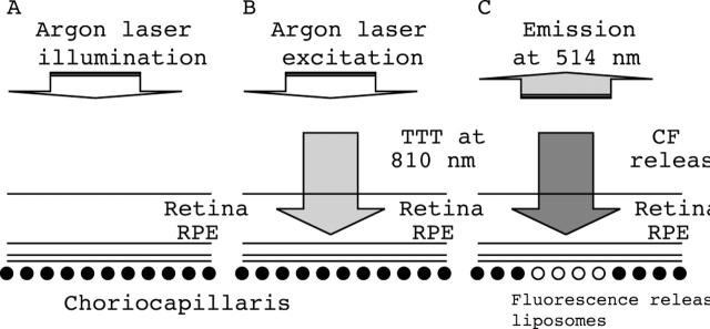
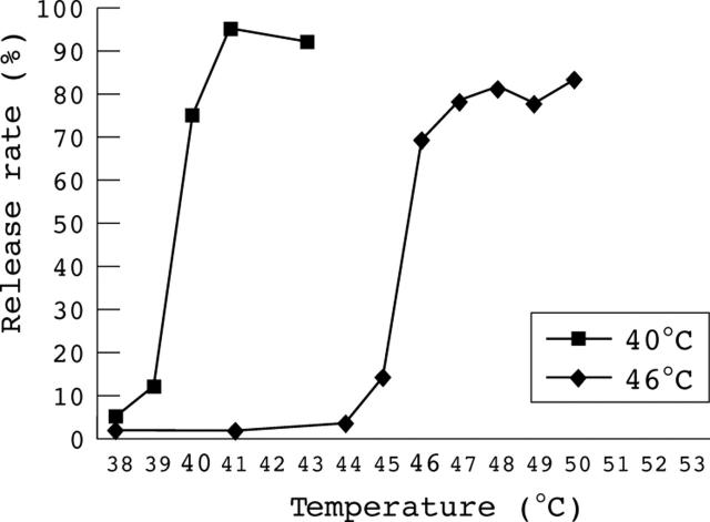
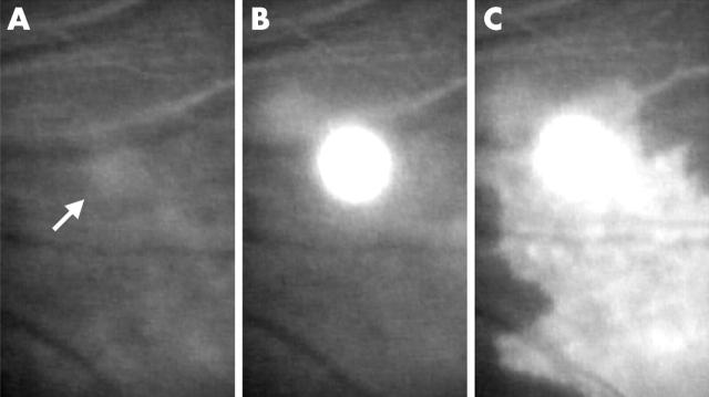
Methods: A modified slit lamp, which was installed with two laser wavelengths (490 nm for illumination and fluorescein excitation and 810 nm for hyperthermia), was developed for TTT and temperature monitoring. Temperature rise during TTT was monitored by observing fluorescence released from thermosensitive liposomes encapsulating carboxyfluorescein. Two types of liposomes were prepared; their phase transition temperatures were 40 degrees C and 46 degrees C, respectively. Laser power settings required to observe fluorescence released from 46 degrees C liposome in normal choroid or CNV were compared. Next, the power settings with 0.5 mm and 0.25 mm spot sizes were compared following administration of 40 degrees C liposome or 46 degrees C liposome.
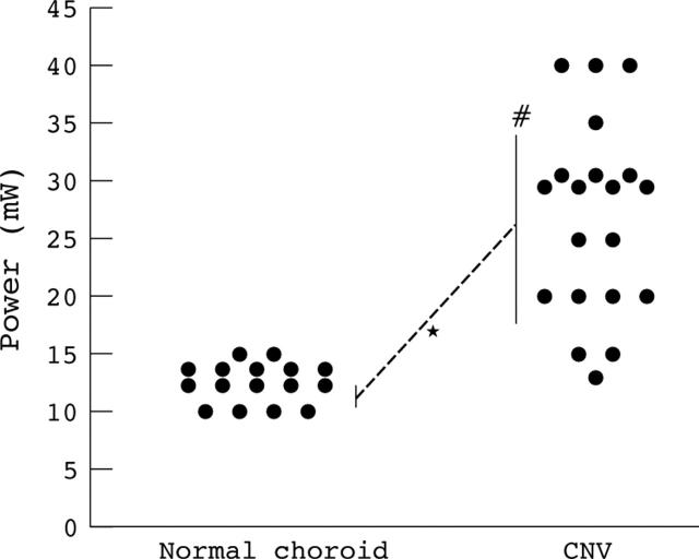
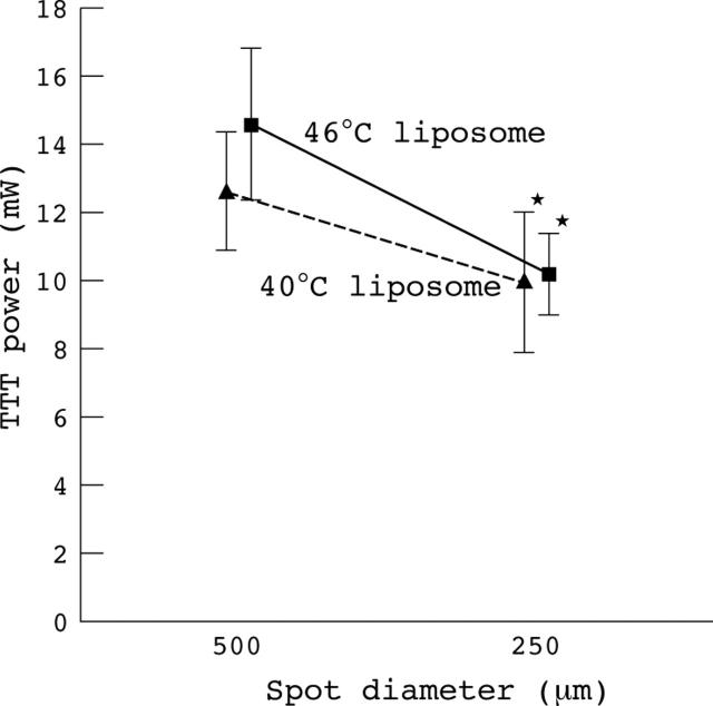
Results: The minimum power values when release from 46 degrees C liposome was observed showed a significant difference in distribution of power values between normal choroid and CNV. CNV required significantly higher power than normal choroid. With 40 degrees C liposome, the power was 9.7 (1.9) mW (mean (SD)) at a spot size of 0.25 mm, and 12.1 (1.6) mW at 0.5 mm, respectively. When using 46 degrees C liposome, the power setting was 10.2 (1.2) mW at a spot size of 0.25 mm, and 14.6 (2.2) mW at 0.5 mm, respectively.
Conclusions: CNV demonstrated varying heat conduction, compared with normal choroid. Laser power required to raise the temperature should not necessarily be doubled, even when the spot size is doubled. Close attention should be given to the selection of power settings when performing TTT for CNV.
Figures
Comment in
-
TTT: local light absorption and heat convection versus heat conduction.Br J Ophthalmol. 2005 Nov;89(11):1545. doi: 10.1136/bjo.2005.082453. Br J Ophthalmol. 2005. PMID: 16234481 Free PMC article. No abstract available.
Similar articles
-
Noninvasive technique for monitoring chorioretinal temperature during transpupillary thermotherapy, with a thermosensitive liposome.Invest Ophthalmol Vis Sci. 2003 Jun;44(6):2716-21. doi: 10.1167/iovs.02-1210. Invest Ophthalmol Vis Sci. 2003. PMID: 12766078
-
Transpupillary thermotherapy for age-related macular degeneration: long-pulse photocoagulation, apoptosis, and heat shock proteins.Ophthalmic Surg Lasers. 2000 Sep-Oct;31(5):359-73. Ophthalmic Surg Lasers. 2000. PMID: 11011704 Review.
-
Retinal function following transpupillary thermotherapy for occult choroidal neovascularization in age-related macular degeneration: a short-term study by focal electroretinography.Acta Ophthalmol Scand. 2006 Feb;84(1):27-35. doi: 10.1111/j.1600-0420.2005.00529.x. Acta Ophthalmol Scand. 2006. PMID: 16445436
-
Subthreshold transpupillary thermotherapy reduces experimental choroidal neovascularization in the mouse without collateral damage to the neural retina.Invest Ophthalmol Vis Sci. 2004 Jun;45(6):1969-74. doi: 10.1167/iovs.03-1329. Invest Ophthalmol Vis Sci. 2004. PMID: 15161865
-
Transpupillary thermotherapy for age-related macular degeneration: principles and techniques.Semin Ophthalmol. 2001 Jun;16(2):55-9. doi: 10.1076/soph.16.2.55.4213. Semin Ophthalmol. 2001. PMID: 15491004 Review.
Cited by
-
TTT: local light absorption and heat convection versus heat conduction.Br J Ophthalmol. 2005 Nov;89(11):1545. doi: 10.1136/bjo.2005.082453. Br J Ophthalmol. 2005. PMID: 16234481 Free PMC article. No abstract available.
-
Which treatment is best for which AMD patient?Br J Ophthalmol. 2006 Feb;90(2):128-30. doi: 10.1136/bjo.2005.083337. Br J Ophthalmol. 2006. PMID: 16424516 Free PMC article.
-
Relationship between intensity of reflected light and temperature increase: assessment of fundus pigmentation for transpupillary thermotherapy.Jpn J Ophthalmol. 2007 Nov-Dec;51(6):462-9. doi: 10.1007/s10384-007-0484-8. Epub 2007 Dec 21. Jpn J Ophthalmol. 2007. PMID: 18158599
References
-
- Freund KB, Yannuzzi LA, Sorenson JA. Age-related macular degeneration and choroidal neovascularization. Am J Ophthalmol 1993;115:786–91. - PubMed
-
- Ferris FL, Fine SL, Hyman L. Age-related macular degeneration and blindness due to neovascular maculopathy. Arch Ophthalmol 1984;102:1640–2. - PubMed
-
- Macular Photocoagulation Study Group. Age-related macular degeneration. The Macular Photocoagulation Study. Am J Ophthalmol 1984;98:376–7. - PubMed
Publication types
MeSH terms
Substances
LinkOut - more resources
Full Text Sources