Comparative Study
. 2005 Apr 6;24(7):1341-51.
doi: 10.1038/sj.emboj.7600628. Epub 2005 Mar 17.

Structural basis of NEDD8 ubiquitin discrimination by the deNEDDylating enzyme NEDP1

Affiliations
Comparative Study

Structural basis of NEDD8 ubiquitin discrimination by the deNEDDylating enzyme NEDP1

Lin-nan Shen et al. EMBO J. .

Abstract

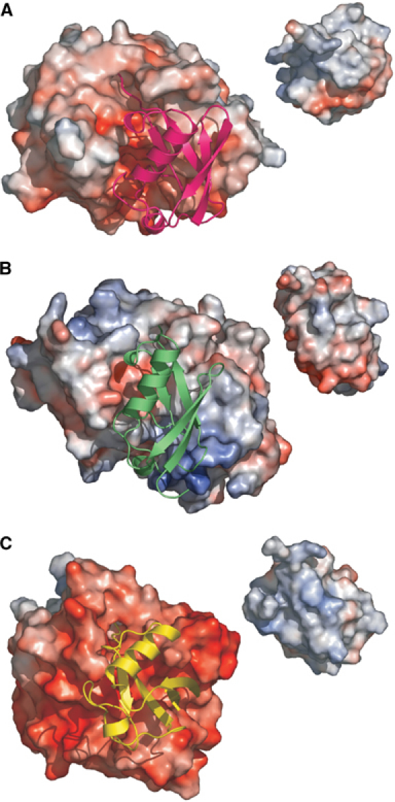
NEDD8 (neural precursor cell expressed developmentally downregulated gene 8)-specific protease NEDP1 processes preNEDD8 to its mature form and deconjugates NEDD8 from substrates such as p53 and cullins. Although NEDD8 and ubiquitin are highly related in sequence and structure, their attachment to a protein leads to different biological effects. It is therefore critical that NEDP1 discriminates between NEDD8 and ubiquitin, and this requires remarkable precision in molecular recognition. To determine the basis of this specificity, we have determined the crystal structure of NEDP1 in isolation and in a transition state complex with NEDD8. This reveals that NEDP1 is a cysteine protease of the Ulp family. Binding of NEDD8 induces a dramatic conformational change in a flexible loop that swings over the C-terminus of NEDD8 locking it into an extended beta-structure optimal for catalysis. Structural, mutational and biochemical studies have identified key residues involved in molecular recognition. A single-residue difference in the C-terminus of NEDD8 and ubiquitin contributes significantly to the ability of NEDP1 to discriminate between them. In vivo analysis indicates that NEDP1 mutants perturb deNEDDylation of the tumour suppressor p53.


Figures

Figure 1
Figure 1
Sequence alignment of NEDD8 with ubiquitin and SUMO and NEDP1 with Senp2 and ULP1. Sequences were aligned using ClustalW (Thompson et al, 1994). Conserved residues are shaded in grey. (A) Sequence alignment of NEDD8 with ubiquitin and SUMO. An alanine marked with an asterisk in NEDD8 was mutated to arginine, while an arginine in ubiquitin was mutated to alanine. Secondary structure elements (in yellow) above the sequence are indicated for NEDD8, while those (blue) below the sequences are indicated for ubiquitin. (B) Sequence alignment of NEDP1 with ULP1 and SENP2. Residues of the catalytic triad are highlighted in red. Residues involved in direct NEDD8 and NEDP1 intermolecular interaction are marked with an asterisk. Mutations in these residues were created by alanine substitution. Residues marked with a triangle were deleted in a loop deletion. The secondary structure elements (yellow) above the sequences are indicated for NEDP1 and those (blue) below the sequences are indicated for SENP2. Sequences shown are from NCBI protein databases: NEDD8 (NP_006147), ubiquitin (AAA36787), SUMO (AAH66306), NEDP1 (AAA36787), SENP2 (AAH40609) and ULP1 (1EUVA).
Figure 2
Figure 2
Structure of NEDP1 alone and in complex with NEDD8. (A) Monomers of NEDP1 from the apo (wheat) and covalent complex (slate). The loop centred around residue 100 is shown in green and is profoundly changed in the complex. (B) The final 2FoFc electron density different map of the linkage contoured at 1σ. (C) Stereo diagram of the NEDP1–NEDD8 complex. NEDP1 is coloured slate and NEDD8 pink. The residues mutated are shown and labelled. (D) The β-sheet-like interaction between nep1 and NEDD8.
Figure 3
Figure 3
Comparison of protease-ubiquitin-like protein complexes. The complex between proteases, shown as electrostatic surfaces with the same scale in each and their target protein shown in a ribbon representation. Inset: The target turned to show its electrostatic surface that binds to the protease. (A) NEDP1–NEDD8. The protease interface is weakly acidic with no basic path. (B) Ulp1–SUMO. The strong positive patch at the bottom and a weaker smaller one at the top of protease are visible. (C) Yuh1–ubiquitin. The protease interface is much more acidic. In comparing ubiqutin and NEDD8, it can be seen that NEDD8 is less basic. This is consistent with changes in residues E53 and A72 (to Gly and Arg respectively in ubiquitin).
Figure 4
Figure 4
Processing activities of NEDP1 mutant proteins. The substrate used in the assay is His-MBP-NEDD8-Ub and the assays are as detailed in Material and methods. NEDP1 and its mutants were mixed with the substrate and after incubation for 30 min at 37°C, the reaction was stopped by adding 6 × Laemmli loading buffer and boiled for 3 min. The processing products were fractionated using SDS–PAGE and the gel stained with 0.25% Coomassie blue. The designation of each mutant enzyme is indicated. CON: substrate only. The upper arrow indicates the substrate His-MBP-NEDD8-Ub, while the lower arrow indicates the cleavage product His-MBP-NEDD8. The released Ub has migrated off the bottom of the gel and is therefore not detected. Data presented are representative of three independent experiments.
Figure 5
Figure 5
Ability of NEDP1 mutants to deconjugate NEDDylated p53 in vivo. Top panel: H1299 cells were transfected with expression constructs for p53, mdm2, 6His-NEDD8 and NEDP1 mutants as indicated. At 36 h after transfection, cells were lysed in guanidine hydrochloride and 6His-NEDD8-conjugated species purified on Ni-NTA agarose as described in Materials and methods. NEDDylated p53 was detected by Western blotting with DO-1 anti-p53 monoclonal antibody. Total levels of NEDP1 are shown in the bottom panel. Middle panel: As in top panel, but 6His-ubiquitin was cotransfected rather than 6His-NEDD8.
Figure 6
Figure 6
How NEDP1 discriminates between NEDD8 and ubiquitin. (A) Superposition of NEDD8 and ubiquitin, the key change is the backbone centred on residue 72. (B) The NEDP1–NEDD8 complex is shown coloured as before. Ubiquitin is shown as an yellow ribbon. The ubiquitin is positioned based on a superposition of NEDP1 and Yuh1. It is clear that ubiquitin has a very different spatial relationship to its protease. Residue 72 is shown and labelled; it is the kink at this position that is responsible for the dramatic difference in orientation. (C) Time course of NEDP1 protein on processing wild-type NEDD8 and NEDD8 A/R mutant. NEDP1 enzyme was incubated with NEDD8 and A72R mutant proteins at 37°C and the reaction was stopped at 0.5, 1, 2, 3, 4 and 6 min by adding an equal volume of 2 × Laemmli loading buffer and boiling for 3 min. The His-MBP-N8 was fractionated by SDS–PAGE and quantified by densitometric analysis using a multianalysis system (Doc-2000, Bio-Rad). The data presented are the mean of three independent experiments. Error bars represent the 95% confidence of the calculated means. (D) Activities of NEDP1 and HAUSP on processing of ubiquitin and R72A ubiquitin. Substrates used in the assay are His-MBP-N8-UB, His-MBP-N8(m)-UB (NEDD8 A72R mutant), HisUB-UB and HisUB9(m)-UB (ubiquitin R72A mutant). Substrates were mixed with NEDP1 or HAUSP and incubated for 30 min at 37°C. The processing products were fractionated in SDS–PAGE and stained with 0.25% Coomassie blue.

References

    1. Brons-Poulsen J, Petersen NE, Horder M, Kristiansen K (1998) An improved PCR-based method for site directed mutagenesis using megaprimers. Mol Cell Probes 12: 345–348 - PubMed
    1. Chiba T, Tanaka K (2004) Cullin-based ubiquitin ligase and its control by NEDD8-conjugating system. Curr Protein Pept Sci 5: 177–184 - PubMed
    1. Collaborative Computational Project N (1994) The CCP4 suite: programs for protein crystallography. Acta Crystallogr D 50: 760–763 - PubMed
    1. Cope GA, Suh GS, Aravind L, Schwarz SE, Zipursky SL, Koonin EV, Deshaies RJ (2002) Role of predicted metalloprotease motif of Jab1/Csn5 in cleavage of Nedd8 from Cul1. Science 298: 608–611 - PubMed
    1. Deshaies RJ (1999) SCF and Cullin/Ring H2-based ubiquitin ligases. Annu Rev Cell Dev Biol 15: 435–467 - PubMed

Publication types

MeSH terms