. 2005 Mar 23;24(6):1146-56.
doi: 10.1038/sj.emboj.7600605. Epub 2005 Mar 3.

E-cadherin is essential for in vivo epidermal barrier function by regulating tight junctions

Affiliations

E-cadherin is essential for in vivo epidermal barrier function by regulating tight junctions

Judith A Tunggal et al. EMBO J. .

Abstract

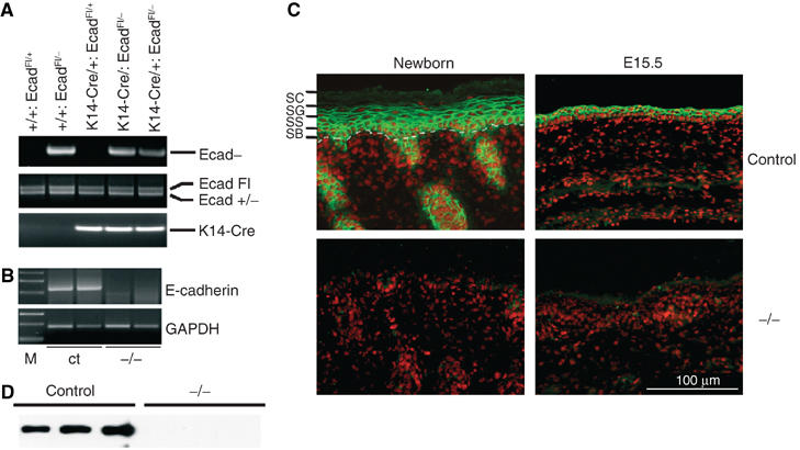
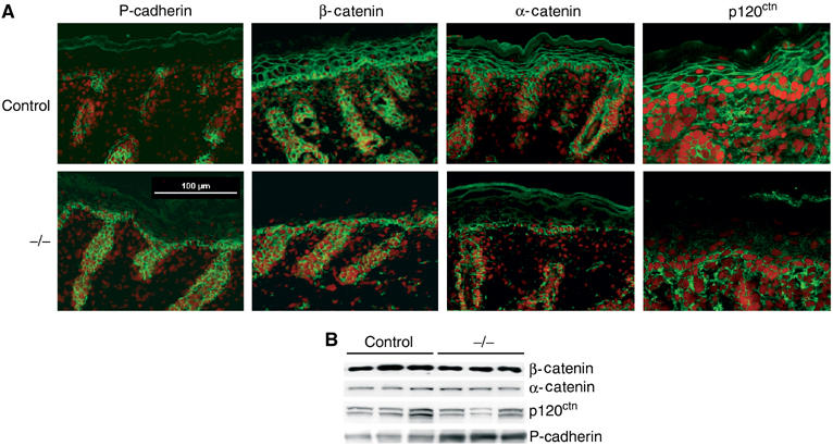
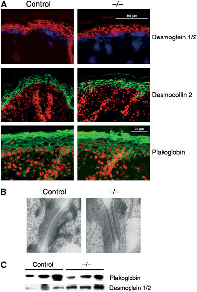
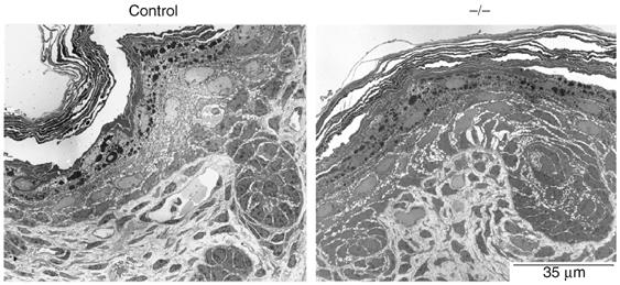
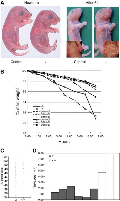
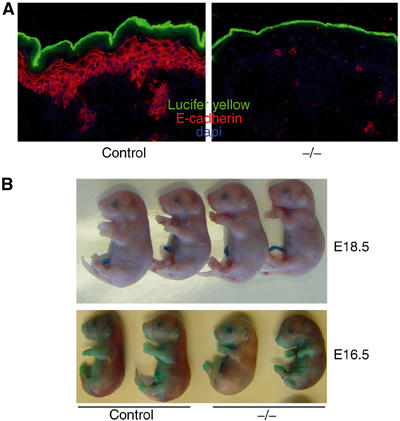
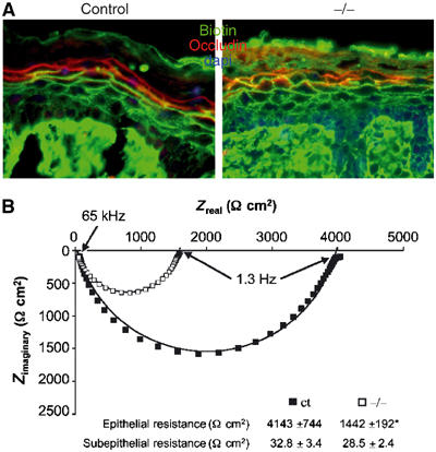
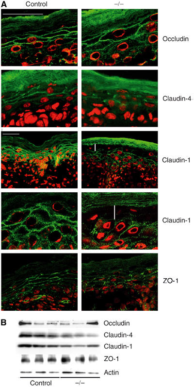
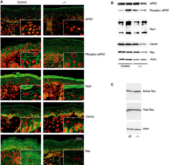
Cadherin adhesion molecules are key determinants of morphogenesis and tissue architecture. Nevertheless, the molecular mechanisms responsible for the morphogenetic contributions of cadherins remain poorly understood in vivo. Besides supporting cell-cell adhesion, cadherins can affect a wide range of cellular functions that include activation of cell signalling pathways, regulation of the cytoskeleton and control of cell polarity. To determine the role of E-cadherin in stratified epithelium of the epidermis, we have conditionally inactivated its gene in mice. Here we show that loss of E-cadherin in the epidermis in vivo results in perinatal death of mice due to the inability to retain a functional epidermal water barrier. Absence of E-cadherin leads to improper localization of key tight junctional proteins, resulting in permeable tight junctions and thus altered epidermal resistance. In addition, both Rac and activated atypical PKC, crucial for tight junction formation, are mislocalized. Surprisingly, our results indicate that E-cadherin is specifically required for tight junction, but not desmosome, formation and this appears to involve signalling rather than cell contact formation.


Figures

Figure 1
Figure 1
(A) PCR analysis of offspring from E-cadherinFl/Fl mice crossed with a deleter K14-Cre/E-cadherin+/− line. Indicated is the DNA fragment generated by PCR reaction. (B) RT–PCR analysis of RNA isolated from skin biopsies of control (ct) and K14-Cre/EcadFl/− (−/−) mice. M: marker. (C) Immunohistochemical analysis of newborn and embryonic day 15.5 (E15.5) skin sections for E-cadherin (green). Nuclei were counterstained with propidium iodide (red). The different epidermal layers are also indicated: SB: stratum basale or basal layer; SS: stratum spinosum or spinal layer; SG: stratum granulosum or granular layer; SC: stratum corneum or cornified layer. The dotted white line indicates the epidermal–dermal junction. (D) Western blot analysis of total newborn mouse skin for E-cadherin.
Figure 2
Figure 2
Adherens junctions expression is altered in K14-Cre/EcadFl/− mice. (A) Immunohistochemical analysis of newborn control and K14-Cre/EcadFl/− (−/−) skin sections stained for adherens junction markers. Nuclei are counterstained with propidium iodide. (B) Western blot analysis of total newborn skin lysates for the indicated adherens junction markers.
Figure 3
Figure 3
Desmosomes are formed in the absence of E-cadherin. (A) Immunohistochemical analysis of newborn control and K14-Cre/EcadFl/− (−/−) skin sections stained for desmosomal markers. Desmoglein 1/2 is red with nuclei counterstained with dapi (blue) whereas desmocollin 2 and plakoglobin are green with nuclei counterstained with propidium iodide (red). (B) Ultrastructural analysis of desmosomes in newborn control and K14-Cre/EcadFl/− (−/−) skin. (C) Western blot analysis of total newborn skin lysates for plakoglobin and desmoglein 1/2.
Figure 4
Figure 4
Normal structure and cell–cell contacts in K14-Cre/EcadFl/− mice. Ultrastructural analysis of newborn control and K14-Cre/EcadFl/− (−/−) skin.
Figure 5
Figure 5
K14-Cre/EcadFl/− mice die of water loss. (A) Macroscopic appearance of newborn control E-cadherin and K14-Cre/EcadFl/− mice. The inset shows close-up of the ventral side. (B) Weight time-course analysis of newborn control and K14-Cre/EcadFl/− mice. (C) Haematocrit analysis of blood isolated from control (n=35) and mutant mice (n=13). (D) TEWL measurements of control and E-cadherin mutant mice.
Figure 6
Figure 6
Epidermal loss of E-cadherin does not affect the outside-in barrier. (A) Lucifer yellow dye penetration assay of newborn control and E-cadherin mutant mice. No penetration of lucifer yellow across the stratum corneum is observed when comparing control mice to K14-Cre/EcadFl/− mice. (B) Toluidine blue penetration assay of E18.5- and E16.5-day-old control and K14-Cre/EcadFl/− mice.
Figure 7
Figure 7
Tight junctions are functionally impaired in K14-Cre/EcadFl/− mice. (A) Inside-out permeability assay. Skin sections of control and mutant newborn mice intradermally injected with biotin were stained with streptavidin to follow the penetration of biotin (green) and counterstained with occludin to mark the tight junctions present in the granular layer. (B) Impedance measurements of ventral skin biopsies isolated from newborn control and E-cadherin mutant mice. Typical experiments are depicted in Nyquist plots; numerical values represent mean±s.e.m. *P-value=0.03. N=8 for control and N=4 for K14-Cre/EcadFl/− (−/−) mice.
Figure 8
Figure 8
Improper formation of tight junctions in K14-Cre/EcadFl/− mice. (A) Immunohistochemical analysis of skin sections for the tight junctional markers (green) occludin, claudin-4, claudin-1 and ZO-1. Nuclei were counterstained with propidium iodide (red). For claudin-1, both × 400 and × 1000 images are shown. White vertical lines indicate absence of claudin-1 staining from granular layer. White bars=25 μM. (B) Western blot of total skin lysates isolated from newborn control and K14-Cre/EcadFl/− (−/−) mice using antibodies against the indicated tight junctional proteins.
Figure 9
Figure 9
Altered localization of activated atypical PKC and the small GTPase Rac in K14-Cre/EcadFl/− mice. (A) Immunohistochemical analysis of skin sections for the indicated polarity complex proteins and the small GTPases Rac and Cdc42 (green). Nuclei were counterstained with propidium iodide (red). (B) Western blot analysis of the indicated proteins in total skin lysates isolated from newborn control and K14-Cre/EcadFl/− (−/−) mice. (C) Rac activation assay on total skin lysates of control and K14-Cre/EcadFl/− mice.

References

    1. Anderson JM, Van Itallie CM, Fanning AS (2004) Setting up a selective barrier at the apical junction complex. Curr Opin Cell Biol 16: 140–145 - PubMed
    1. Bilder D, Schober M, Perrimon N (2003) Integrated activity of PDZ protein complexes regulates epithelial polarity. Nat Cell Biol 5: 53–58 - PubMed
    1. Boussadia O, Kutsch S, Hierholzer A, Delmas V, Kemler R (2002) E-cadherin is a survival factor for the lactating mouse mammary gland. Mech Dev 115: 53–62 - PubMed
    1. Braga VM (2002) Cell–cell adhesion and signalling. Curr Opin Cell Biol 14: 546–556 - PubMed
    1. Brandner JM, Kief S, Grund C, Rendl M, Houdek P, Kuhn C, Tschachler E, Franke WW, Moll I (2002) Organization and formation of the tight junction system in human epidermis and cultured keratinocytes. Eur J Cell Biol 81: 253–263 - PubMed

Publication types

MeSH terms