Comparative Study
. 2005 Apr;6(4):354-60.
doi: 10.1038/sj.embor.7400366.

Variant histone H3.3 marks promoters of transcriptionally active genes during mammalian cell division

Affiliations
Comparative Study

Variant histone H3.3 marks promoters of transcriptionally active genes during mammalian cell division

Cheok-Man Chow et al. EMBO Rep. 2005 Apr.

Abstract

Variant histone H3.3 is incorporated into nucleosomes by a mechanism that does not require DNA replication and has also been implicated as a potential mediator of epigenetic memory of active transcriptional states. In this study, we have used chromatin immunoprecipitation analysis to show that H3.3 is found mainly at the promoters of transcriptionally active genes. We also show that H3.3 combines with H3 acetylation and K4 methylation to form a stable mark that persists during mitosis. Our results suggest that H3.3 is deposited principally through the action of chromatin-remodelling complexes associated with transcriptional initiation, with deposition mediated by RNA polymerase II elongation having only a minor role.


Figures

Figure 1
Figure 1
Location of variant histone H3.3 in the endogenous λ5-VpreB1 locus. (A) The λ5-VpreB1 locus. (B) Expression of Myc–H3.3 in stably transfected clones detected using an anti-c-Myc tag antibody (green, left panel). DNA was counterstained with TOTO (blue, right panel). (C) Chromatin immunoprecipitation analysis of H3.3. The positions of the primer pairs are shown on the locus map (below the graph) as vertical bars. DNase I-hypersensitive sites (HS) that have been mapped in the locus are shown as vertical arrows (red, constitutive HS; black, pre-B-cellspecific HS). Enrichment values represent the mean±s.d. from two independently immunoprecipitated samples (see Methods for details of calculations).
Figure 2
Figure 2
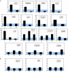
Comparison of the levels of Myc–H3.3 in the 5′ promoter regions and the transcribed regions of different genes. Results were obtained by chromatin immunoprecipitation analysis of Myc–H3.3-expressing pre-B cells using anti-c-Myc tag antibody. PCR values were used to calculate enrichment relative to non-transfected cells (see Methods). Because the values are calculated as ratios relative to non-transfected cells, values ⩽1 (blue shading) indicate no enrichment (1) or depletion (<1). For PCR primer positions, see supplementary Table 1 online. For analysis of transcription levels of the genes, see supplementary Fig 1 online. (A) Genes that are transcribed in pre-B cells. The significance of the differences between the promoters and the transcribed regions was tested for the entire transcribed gene data set using the non-parametric Wilcoxon's matched-pairs test (P=0.01). (B) Genes that are silent in pre-B cells. p, promoter; t, transcribed region; td, distal transcribed region; tp, proximal transcribed region.
Figure 3
Figure 3
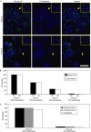
Localization of variant histone H3.3 to a variegating λ5 transgene on metaphase chromosomes. (A) The λ5 transgene. (B) RNA fluorescence in situ hybridization (FISH) analysis of a variegated pre-B-cell clone showing expression of the λ5 transgene (green) and endogenous β-actin (red). Scale bar, 10 μm. (C) Immuno-DNA FISH analysis showing colocalization of Myc–H3.3 (green) and the λ5 transgene (red) on metaphase chromosomes in a clone with greater than 70% of cells expressing the transgene (clone 1) and one with less than 2% of cells expressing the transgene (clone 2). DNA was counterstained with TOTO (blue). Immuno-FISH was also carried out on metaphase spreads from clone 1 using antibodies specific for phosphorylated histone H3 (green, bottom panel). Arrows indicate the pericentromeric region. Scale bar, 2 μm. (D) Comparison of Myc–H3.3 colocalization with the transgene during mitosis with numbers of expressing cells in two different pre-B-cell clones. Confocal images of metaphase spreads were randomized and blindscored by an operator not involved in the analysis. The number of cells or metaphase spreads analysed for each clone is indicated below each histogram bar. χ2 analysis of the values showed that the differences between the clones were highly significant (P<0.0001).
Figure 4
Figure 4
Localization of acetylated and methylated histone H3 to a variegating λ5 transgene on metaphase chromosomes. (A) Immunofluorescence in situ hybridization (Immuno-FISH) analysis showing colocalization of diacetylated histone H3 (green) and the λ5 transgene (red) during mitosis in a clone with greater than 90% expressing cells (clone 3) and one with less than 5% expressing cells (clone 6). DNA was counterstained with TOTO (blue). Arrows indicate the position of the transgene. Scale bar, 10 μm. The insets show the chromosome containing the transgene at higher magnification. (B) Correlation of histone H3 acetylation at the transgene with numbers of expressing cells in four different pre-B-cell clones. The number of cells or metaphases analysed for each clone is indicated below each histogram bar. Correlation analysis is shown in supplementary Fig 5B online (R=0.99). (C) Immuno-DNA FISH analysis of the transgene on metaphase chromosomes using antibodies specific for the di- or trimethylated form of H3 K4. χ2 analysis showed that the difference between the clones was highly significant (P<0.01).

References

    1. Agalioti T, Chen G, Thanos D (2002) Deciphering the transcriptional histone acetylation code for a human gene. Cell 111: 381–392 - PubMed
    1. Ahmad K, Henikoff S (2002) The histone variant H3.3 marks active chromatin by repliation-independent nucleosome assembly. Mol Cell 9: 1191–1200 - PubMed
    1. Akhmanova A, Miedema K, Wang Y, van Bruggen M, Berden JH, Moudrianakis EN, Hennig W (1997) The localization of histone H3.3 in germ line chromatin of Drosophila males as established with a histone H3.3specific antiserum. Chromosoma 106: 335–347 - PubMed
    1. Christova R, Oelgeschlager T (2002) Association of human TFIID-promoter complexes with silenced mitotic chromatin in vivo. Nat Cell Biol 4: 79–82 - PubMed
    1. Henikoff S, Furuyama T, Ahmad K (2004) Histone variants, nucleosome assembly and epigenetic inheritance. Trends Genet 20: 320–326 - PubMed

Publication types

MeSH terms