. 2005 Apr;115(4):959-68.
doi: 10.1172/JCI19935. Epub 2005 Mar 17.

Cholesterol targeting alters lipid raft composition and cell survival in prostate cancer cells and xenografts

Affiliations

Cholesterol targeting alters lipid raft composition and cell survival in prostate cancer cells and xenografts

Liyan Zhuang et al. J Clin Invest. 2005 Apr.

Abstract

Lipid rafts are cholesterol- and sphingolipid-enriched microdomains in cell membranes that regulate phosphorylation cascades originating from membrane-bound proteins. In this study, we tested whether alteration of the cholesterol content of lipid rafts in prostate cancer (PCa) cell membranes affects cell survival mechanisms in vitro and in vivo. Simvastatin, a cholesterol synthesis inhibitor, lowered raft cholesterol content, inhibited Akt1 serine-threonine kinase (protein kinase Balpha)/protein kinase B (Akt/PKB) pathway signaling, and induced apoptosis in caveolin- and PTEN-negative LNCaP PCa cells. Replenishing cell membranes with cholesterol reversed these inhibitory and apoptotic effects. Cholesterol also potentiated Akt activation in normal prostate epithelial cells, which were resistant to the apoptotic effects of simvastatin. Elevation of circulating cholesterol in SCID mice increased the cholesterol content and the extent of protein tyrosine phosphorylation in lipid rafts isolated from LNCaP/sHB xenograft tumors. Cholesterol elevation also promoted tumor growth, increased phosphorylation of Akt, and reduced apoptosis in the xenografts. Our results implicate membrane cholesterol in Akt signaling in both normal and malignant cells and provide evidence that PCa cells can become dependent on a cholesterol-regulated Akt pathway for cell survival.


Figures

Figure 1
Figure 1
Simvastatin treatment downregulates Akt phosphorylation and Akt kinase activity and induces apoptosis in LNCaP cells. (A) Cells were incubated with varying doses of simvastatin (sim) in the absence or presence of cholesterol complexes for 16 hours. Whole-cell lysates were resolved by SDS-PAGE and immunoblotted with antibodies to total Akt or S473-phosphorylated Akt (S473-P). (B) LNCaP cells were incubated in the presence of 20 μM simvastatin or vehicle (control) in serum-free medium at 37°C for the indicated times, after which lysates were collected for Western blot and in vitro kinase assay. A GSK3 fusion protein was used as Akt substrate after immunoprecipitation with anti-Akt antibody. Kinase assay eluates were blotted with antibodies to total Akt, phospho-GSK3α/β (p-GSK3α/β), T308-P Akt, and S473-P Akt. (C) Cells in serum-free medium were treated with 20 μM simvastatin in the absence or presence of cholesterol (chol) complexes at 37°C for 12 hours, after which lysates were collected and kinase assay was performed as in B. (D) LNCaP cells were treated for varying times with 20 μM simvastatin. Apoptosis was determined by DNA fragmentation. The means ± SD of triplicate determinations are shown. (E) LNCaP cells were treated with 20 μM simvastatin with or without cholesterol complexes for 12 hours followed by DNA fragmentation analysis (*P < 0.05). (F) LNCaP cells were incubated with 20 μM simvastatin with or without cholesterol complexes for 12 hours, after which lysates were collected for immunoblot with the indicated antibodies. c-Caspase-7, cleaved caspase-7.
Figure 2
Figure 2
Simvastatin treatment reduces the cholesterol content of lipid rafts of LNCaP cells and inhibits Akt phosphorylation in rafts. (A) Immunoblot results obtained following fractionation of Triton X-100–insoluble material by sucrose gradient ultracentrifugation. This panel demonstrates how lipid raft fractions used for the cholesterol determinations shown in B were obtained. Flotation fractions demonstrating enrichment in the raft markers Giα2 and flotillin-2 (i.e., fraction 6 in this example) were designated as raft fractions. (B) Cells were incubated in serum-free medium in the absence (control) or presence of 10 μM simvastatin overnight at 37°C. After the drug treatment, 1 group was incubated with cholesterol complexes (sim + chol) at 37°C for 1 hour. The cholesterol/protein ratio was determined in lipid raft fractions prepared as shown in A and under the conditions described in the text. Values shown are means ± SD of triplicate determinations (*P < 0.01). (C) Cells were incubated in the presence of 20 μM simvastatin or vehicle in serum-free medium at 37°C for the indicated times. C+M and raft fractions were isolated by successive detergent extraction, resolved by SDS-PAGE, transferred to nitrocellulose membranes, and immunoblotted with the indicated antibodies. (D) Cells were treated with 20 μM simvastatin with or without cholesterol complexes for 4 hours, followed by raft extraction and analysis as in C.
Figure 3
Figure 3
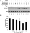
Cholesterol depletion inhibits Akt phosphorylation but does not induce apoptosis in PrECs. (A) Immunodetection of Akt in cell lysates following treatments. PrECs were treated with 20 ng/ml EGF, in the presence or absence of cholesterol complexes, under the following conditions: 1 hour of pretreatment with 2 μg/ml filipin (lane 2), 1 hour of pretreatment with 20 mM cyclodextrin (lane 3), 16 hours of pretreatment with 20 μM simvastatin (lane 4), 1 hour of pretreatment with vehicle (lane 5). After 1 hour of cholesterol pretreatment (lane 6), some groups were incubated with 2 μg/ml filipin (lane 7) or 20 mM cyclodextrin (lane 8). Other groups treated identically to conditions in lanes 7 and 8 were repleted (asterisk) with cholesterol for 1 hour (lanes 9 and 10). (B) PrECs were treated with varying concentrations of simvastatin or vehicle for 24 hours, and the extent of apoptosis was determined by DNA fragmentation. Values shown are means ± SD of triplicate determinations.
Figure 4
Figure 4
High levels of serum cholesterol increase tumor aggressiveness. (A) Serum cholesterol levels in venous blood after stable elevation using dietary modification for 4 weeks. Values are means ± SD of determinations from 5 animals (P < 0.001). (B) Subcutaneous xenograft tumors were created by subcutaneous injection of LNCaP/sHB cells after stable cholesterol elevation was demonstrated. The tumor take was significantly different between normal and high-cholesterol groups (P < 0.0001). (C) Mice were sacrificed 6 weeks after tumor cell injection. Four representative xenograft tumors from each group are shown. (D) Volume measurements were made at 5 weeks and 6 weeks after tumor cell injection. Median tumor volumes (horizontal lines) for the normal group were 0.077 cm3 (5 weeks) and 0.099 cm3 (6 weeks); median tumor volumes for the hypercholesteremic group were 0.135 cm3 (5 weeks) and 0.141 cm3 (6 weeks) (*P < 0.01; **P < 0.005).
Figure 5
Figure 5
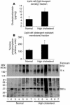
Elevated cholesterol and protein tyrosine phosphorylation in lipid rafts isolated from xenograft tumors exposed to high circulating cholesterol. (A) Cholesterol level in lipid rafts isolated from LNCaP/sHB xenograft tumors by sucrose density gradient ultracentrifugation (n = 4 tumors per condition) (P < 0.01). (B) Evidence for increased tyrosine phosphorylation of lipid raft proteins isolated from LNCaP/sHB xenograft tumors by differential solubilization in Triton X-100 and OCG (38). OCG-soluble proteins were resolved by SDS-PAGE, transferred to nitrocellulose, and blotted with anti-phosphotyrosine antibody (right panel). Top panel: Quantitative evaluation by scanning densitometry of immunoblot shown on the bottom (P < 0.0005). MW, molecular weight.
Figure 6
Figure 6
Increased Akt activation in xenograft tumors from mice with high circulating cholesterol. (AC) Tests of S473-P antibody specificity for the experiment shown in D. LNCaP cells were starved in serum-free medium at 37°C for 16 hours and then treated with 0.5 mM pervanadate for 15 minutes at 37°C. Original magnification: A, ×200; B and C, ×600. (D) Anti–phospho-Akt1 antibody was used to detect the status of Akt activation in LNCaP/sHB xenograft tumors from normal and high-cholesterol animals with the immunofluorescence procedures used for AC. Two representative images are shown (original magnification, ×400). Optical intensities of the images were determined automatically with computer-controlled software (n = 4 in each group). The values shown are mean signal intensity (INT) per square millimeter ± SD versus group (P < 0.001).
Figure 7
Figure 7
Reduced apoptosis in xenograft tumors from mice with high circulating cholesterol. Apoptosis rates in LNCaP/sHB xenograft tumors from normal (n = 3) and high-cholesterol (n = 4) groups as evaluated by TUNEL (original magnification, ×200). Fluorescence originates from condensed nuclei in apoptotic cells. The graph is presented as percent apoptotic cells (apoptotic cells/total cells) ± SD versus group (P < 0.001).

Similar articles

Cited by

References

    1. Pike LJ. Lipid rafts: bringing order to chaos. J. Lipid Res. 2003;44:655–667. - PubMed
    1. Deurs B, Roepstorff K, Hommelgaard AM, Sandvig K. Caveolae: anchored, multifunctional platforms in the lipid ocean. Trends Cell Biol. 2003;13:92–100. - PubMed
    1. Drab M, et al. Loss of caveolae, vascular dysfunction, and pulmonary defects in caveolin-1 gene-disrupted mice. Science. 2001;293:2449–2452. - PubMed
    1. Park DS, et al. Caveolin-1/3 double-knockout mice are viable, but lack both muscle and non-muscle caveolae, and develop a severe cardiomyo-pathic phenotype. Am. J. Pathol. 2002;160:2207–2217. - PMC - PubMed
    1. Liu P, Rudick M, Anderson RG. Multiple functions of caveolin-1. J. Biol. Chem. 2002;277:41295–41298. - PubMed

Publication types

MeSH terms