. 2005 Apr 10;334(2):306-18.
doi: 10.1016/j.virol.2005.02.001.

Spike protein assembly into the coronavirion: exploring the limits of its sequence requirements

Affiliations

Spike protein assembly into the coronavirion: exploring the limits of its sequence requirements

Berend Jan Bosch et al. Virology. .

Abstract

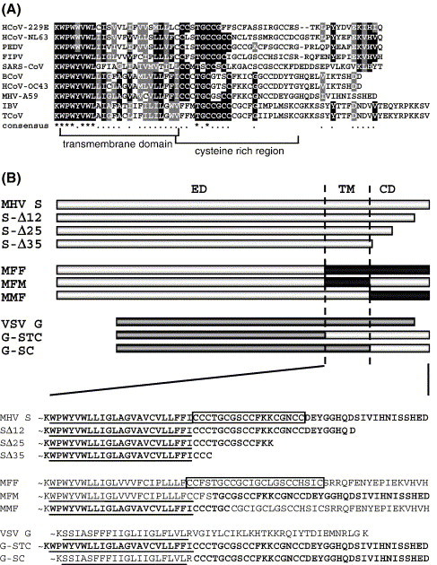
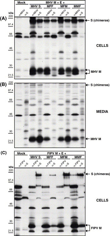
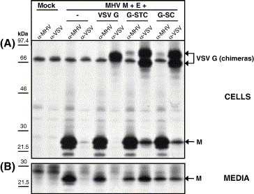
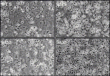
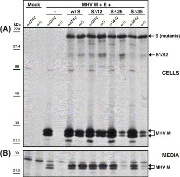
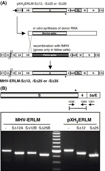
The coronavirus spike (S) protein, required for receptor binding and membrane fusion, is incorporated into the assembling virion by interactions with the viral membrane (M) protein. Earlier we showed that the ectodomain of the S protein is not involved in this process. Here we further defined the requirements of the S protein for virion incorporation. We show that the cytoplasmic domain, not the transmembrane domain, determines the association with the M protein and suffices to effect the incorporation into viral particles of chimeric spikes as well as of foreign viral glycoproteins. The essential sequence was mapped to the membrane-proximal region of the cytoplasmic domain, which is also known to be of critical importance for the fusion function of the S protein. Consistently, only short C-terminal truncations of the S protein were tolerated when introduced into the virus by targeted recombination. The important role of the about 38-residues cytoplasmic domain in the assembly of and membrane fusion by this approximately 1300 amino acids long protein is discussed.


Figures

Fig. 1
Fig. 1
(A) A CLUSTALW alignment of the carboxy-terminal spike protein sequences from nine coronaviruses including feline infectious peritonitis virus (FIPV, strain 79-1146; GenBank accession no. VGIH79), porcine transmissible gastroenteritis virus (TGEV, strain Purdue; GenBank accession no. P07946), porcine epidemic diarrhea virus (PEDV; GenBank accession no. NP_598310), HCoV-229E (human coronavirus, strain 229E; GenBank accession no. VGIHHC), bovine coronavirus (BcoV, strain F15; GenBank accession no. P25190), mouse hepatitis virus (MHV, strain A59; GenBank accession no. P11224), HCoV-OC43 (human coronavirus, strain OC43; GenBank accession no. CAA83661), SARS-CoV (strain TOR2; GenBank accession no. P59594) and infectious bronchitis virus (IBV, strain Beaudette; GenBank accession no. P11223). The transmembrane domain and cysteine-rich region are indicated. (B) Diagrams of wild-type MHV S protein; cytoplasmic domain truncation mutants SΔ12, SΔ25, and SΔ35; MHV-FIPV chimeras MFF, MFM, and MMF; and VSV G wild-type and VSV-MHV chimeric proteins G-STC and G-SC. Light grey boxes represent the MHV S amino acids sequences, black boxes indicate FIPV S sequences, whereas the dark grey bars represent sequences of the VSV G. ED, ectodomain; TM, transmembrane domain; CD, cytoplasmic domain. Below, TM and CD amino acid sequences of all constructs shown above. The cysteine-rich region in the MHV S and MFF protein has been boxed. The MHV S-derived sequences are indicated in bold. TM regions are underlined.
Fig. 2
Fig. 2
Interactions of the wt MHV S protein or the MHV/FIPV S chimeric proteins MFF, MFM, and MMF with the membrane protein M and their incorporation into VLPs. (A) Demonstration of intracellular interaction of the S protein (chimeras) with M. Intracellular expression of the MHV M and E proteins in combination with the MHV S, MFF, MFM, or MMF protein. Radiolabeled proteins were immunoprecipitated from the cell lysate using the anti-MHV serum (α-MHV) or the anti-S monoclonal antibody (α-S) and analyzed by SDS–PAGE. (B) Demonstration of (chimeric) S protein incorporation into VLPs. Culture media were collected, processed for affinity isolation of radiolabeled VLPs using the anti-MHV serum (α-MHV) or the anti-S monoclonal antibody (α-S), and the samples were analyzed by SDS–PAGE. The molecular mass markers are indicated on the left. Arrows on the right indicate the positions of the expressed proteins. (C) Interactions of the (chimeric) spike protein with the FIPV membrane protein M. Intracellular expression of the FIPV M and E proteins in combination with the MHV S, MFF, MFM, or MMF protein. Radiolabeled proteins were immunoprecipitated from the cell lysate using the anti-FIPV serum (α-FIPV) or the monoclonal antibody to MHV S (α-S) and analyzed by SDS– PAGE.
Fig. 3
Fig. 3
Interactions of the wt VSV G or the VSV-G/MHV-S chimeric proteins G-STC and G-SC with the membrane protein M and their incorporation into VLPs. (A) Demonstration of intracellular interaction of the (chimeric) VSV G with M. Intracellular expression of the MHV M and E proteins in combination with the VSV G, G-STC, and G-SC protein. Radiolabeled proteins were immunoprecipitated from the cell lysate using the anti-MHV serum (α-MHV) or the anti-S monoclonal antibody (α-S) and analyzed by SDS–PAGE. (B) Demonstration of (chimeric) S protein incorporation into VLPs. Culture media were collected, processed for affinity isolation of radiolabeled VLPs using the anti-MHV serum (α-MHV) or the anti-S monoclonal antibody (α-S), and the samples were analyzed by SDS–PAGE. The molecular mass markers are indicated on the left. Arrows on the right indicate the positions of the expressed proteins.
Fig. 4
Fig. 4
Fusion properties of the recombinant spike proteins containing CD truncations. Subconfluent monolayers of OST7-1 cells were infected with vTF7.3 and transfected with the plasmids encoding MHV S protein and the recombinant SΔ12, SΔ25, and SΔ35 CD truncation proteins. At 6 h pi the cells were overlaid with LR7 cells and at 9 h pi pictures were taken.
Fig. 5
Fig. 5
Interactions of the wt MHV S protein or CD truncation proteins SΔ12, SΔ25, and SΔ35 with the membrane protein M and their incorporation into VLPs. (A) Demonstration of intracellular interaction of the S proteins with M. Intracellular expression of the MHV M and E proteins in combination with the wt MHV S or SΔ12, SΔ25, and SΔ35 spike CD truncation proteins. Radiolabeled proteins were immunoprecipitated from the cell lysate using the anti-MHV serum (α-MHV) or the anti-S monoclonal antibody (α-S) and analyzed by SDS–PAGE. (B) Demonstration of S protein incorporation into VLPs. Culture media were collected, processed for affinity isolation of radiolabeled VLPs using the anti-MHV serum (α-MHV) or the anti-S monoclonal antibody (α-S), and the samples were analyzed by SDS–PAGE. The molecular mass markers are indicated on the left. Arrows on the right indicate the positions of the expressed proteins.
Fig. 6
Fig. 6
(A) Plasmid constructs, targeted recombination, and recombinant viruses. The plasmids pXH2ERLM-SΔ12, -SΔ25, and -SΔ35 (see Materials and methods) were used to transcribe the defective RNAs in vitro by using T7 polymerase. The arrow at the left end of the vectors indicates the T7 promoter; the solid circle represents the polylinker between the 5′-end segment of the MHV genome (labeled 5′/1) and the HE gene, which is followed by the structural and group specific genes, the inserted Renilla luciferase gene (RL), the 3′ untranslated region (UTR), and the polyadenylate segment (labeled 3′/U). The asterisk indicates the position of the S protein cytoplasmic domain truncations. The lower part shows a scheme for targeted recombination by using the interspecies chimeric fMHV, which grows only in feline cells. Recombinant viruses generated by the indicated crossover event can be selected on the basis of their ability to grow in murine cells and by the acquired Renilla luciferase gene. (B) RT-PCR analysis of recombinant MHV-ERLM viruses with S protein CD truncations. An (RT-)PCR was used to amplify regions of cytoplasmic viral RNA isolated from cells infected with MHV-ERLM, MHV-ERLM-SΔ12 (clones A and B), or MHV-ERLM-SΔ25 (clone B). The approximate locations of primers 1036, 1090, and 1261 in the recombinant MHV genomes are shown. Primer 1261 was used for the RT-step. Primer pair 1036–1090 was used for the PCR on the RT product and, as a control, on the plasmids used to make the recombinant viruses (pXH2ERLM, -SΔ12, and -SΔ25). PCR products were analyzed in an agarose gel. The most intense band of the 100-bp marker represents the 600 bp DNA. The asterisk marks the position of the S protein cytoplasmic domain truncations.
Fig. 7
Fig. 7
Growth characteristics and sequence analysis of recombinant viruses containing S protein CD truncations. (A) Plaque sizes of MHV-ERLM-SΔ12 clones A and B, MHV-ERLM-SΔ25 clone B, MHV-ERLM-SΔ25 revertant, and MHV-ERLM-SΔ25R clones A and B relative to MHV-ERLM. (B) Single-step growth kinetics of MHV-ERLM-SΔ12 compared to MHV-ERLM. LR7 cells were infected with either MHV-ERLM or MHV-ERLM-SΔ12 clone A or B at an MOI of 5. Viral infectivities in the culture media at different times post-infection were determined by a quantal assay on LR7 cells, and the TCID50 values were calculated. (C) Luciferase detection in infected cell lysates during single-step growth of MHV-ERLM-SΔ12 clones A and B compared to MHV-ERLM. (D) Sequence analysis of MHV-ERLM-SΔ25 revertant viruses. A 17-nucleotide deletion indicated by a dashed line was observed in the sequence of two MHV-ERLM-SΔ25 recombinant viruses that had been passaged independently for 6 rounds and had regained fitness. The deletion resulted in an extension of the cytoplasmic domain with 6 amino acids (SΔ25R) compared to the original MHV-ERLM-SΔ25 recombinant (SΔ25). The translated amino acid sequences corresponding to the S open reading frame of the SΔ25 and SΔ25R viruses are indicated above and below the nucleotide sequences, respectively. Stop codons are marked in bold and by an asterisk.

Similar articles

Cited by

References

    1. Ali A., Nayak D.P. Assembly of Sendai virus: M protein interacts with F and HN proteins and with the cytoplasmic tail and transmembrane domain of F protein. Virology. 2000;276(2):289–303. - PubMed
    1. Bos E.C., Heijnen L., Luytjes W., Spaan W.J. Mutational analysis of the murine coronavirus spike protein: effect on cell-to-cell fusion. Virology. 1995;214(2):453–463. - PMC - PubMed
    1. Cathomen T., Naim H.Y., Cattaneo R. Measles viruses with altered envelope protein cytoplasmic tails gain cell fusion competence. J. Virol. 1998;72(2):1224–1234. - PMC - PubMed
    1. Cavanagh D. The coronavirus surface glycoprotein. In: Siddell S.G., editor. The coronaviridae. Plenum Press; New York: 1995.
    1. Chang K.W., Sheng Y., Gombold J.L. Coronavirus-induced membrane fusion requires the cysteine-rich domain in the spike protein. Virology. 2000;269(1):212–224. - PMC - PubMed

Publication types

MeSH terms

LinkOut - more resources