

Aconitase and ATP synthase are targets of malondialdehyde modification and undergo an age-related decrease in activity in mouse heart mitochondria
- PMID: 15781244
- PMCID: PMC2837075
- DOI: 10.1016/j.bbrc.2005.02.135
Aconitase and ATP synthase are targets of malondialdehyde modification and undergo an age-related decrease in activity in mouse heart mitochondria
Abstract
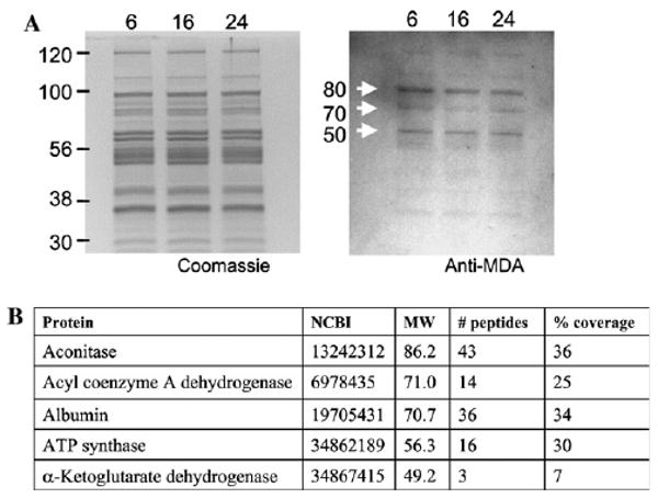
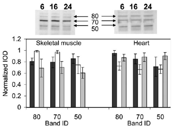
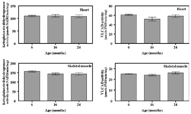
The main purpose of this study was to identify mitochondrial proteins that exhibit post-translational oxidative modifications during the aging process and to determine the resulting functional alterations. Proteins forming adducts with malondialdehyde (MDA), a product of lipid peroxidation, were identified by immunodetection in mitochondria isolated from heart and hind leg skeletal muscle of 6-, 16-, and 24-month-old mice. Aconitase, very long chain acyl coenzyme A dehydrogenase, ATP synthase, and alpha-ketoglutarate dehydrogenase were detected as putative targets of oxidative modification by MDA. Aconitase and ATP synthase from heart exhibited significant decreases in activity with age. Very long chain acyl coenzyme A dehydrogenase and alpha-ketoglutarate dehydrogenase activities were unaffected during aging in both heart and skeletal muscle. This suggests that the presence of a post-translational oxidative modification in a protein does not a priori reflect an alteration in activity. The biological consequences of an age-related decrease in aconitase and ATP synthase activities may contribute to the decline in mitochondrial bioenergetics evident during aging.
Figures

Similar articles
-
Posttranslational modifications and dysfunction of mitochondrial enzymes in human heart failure.Am J Physiol Endocrinol Metab. 2016 Aug 1;311(2):E449-60. doi: 10.1152/ajpendo.00127.2016. Epub 2016 Jul 12. Am J Physiol Endocrinol Metab. 2016. PMID: 27406740
-
Aconitase is the main functional target of aging in the citric acid cycle of kidney mitochondria from mice.Mech Ageing Dev. 2006 Jan;127(1):79-84. doi: 10.1016/j.mad.2005.09.028. Epub 2005 Nov 10. Mech Ageing Dev. 2006. PMID: 16289253 Free PMC article.
-
Age-related impairment of mitochondrial matrix aconitase and ATP-stimulated protease in rat liver and heart.Eur J Biochem. 2004 Nov;271(22):4559-64. doi: 10.1111/j.1432-1033.2004.04422.x. Eur J Biochem. 2004. PMID: 15560797
-
Post-translational modifications of ATP synthase in the heart: biology and function.J Bioenerg Biomembr. 2009 Apr;41(2):145-50. doi: 10.1007/s10863-009-9218-6. J Bioenerg Biomembr. 2009. PMID: 19399597 Free PMC article. Review.
-
Aconitase post-translational modification as a key in linkage between Krebs cycle, iron homeostasis, redox signaling, and metabolism of reactive oxygen species.Redox Rep. 2014 Jan;19(1):8-15. doi: 10.1179/1351000213Y.0000000073. Epub 2013 Nov 22. Redox Rep. 2014. PMID: 24266943 Free PMC article. Review.
Cited by
-
4-Hydroxy-2-nonenal, a reactive product of lipid peroxidation, and neurodegenerative diseases: a toxic combination illuminated by redox proteomics studies.Antioxid Redox Signal. 2012 Dec 1;17(11):1590-609. doi: 10.1089/ars.2011.4406. Epub 2012 Feb 15. Antioxid Redox Signal. 2012. PMID: 22114878 Free PMC article. Review.
-
Redox proteomics in selected neurodegenerative disorders: from its infancy to future applications.Antioxid Redox Signal. 2012 Dec 1;17(11):1610-55. doi: 10.1089/ars.2011.4109. Epub 2012 Jan 18. Antioxid Redox Signal. 2012. PMID: 22115501 Free PMC article. Review.
-
Skeletal muscle mitochondria and aging: a review.J Aging Res. 2012;2012:194821. doi: 10.1155/2012/194821. Epub 2012 Jul 19. J Aging Res. 2012. PMID: 22888430 Free PMC article.
-
The Advanced Lipoxidation End-Product Malondialdehyde-Lysine in Aging and Longevity.Antioxidants (Basel). 2020 Nov 15;9(11):1132. doi: 10.3390/antiox9111132. Antioxidants (Basel). 2020. PMID: 33203089 Free PMC article. Review.
-
Mitochondrial Aconitase and Its Contribution to the Pathogenesis of Neurodegenerative Diseases.Int J Mol Sci. 2024 Sep 15;25(18):9950. doi: 10.3390/ijms25189950. Int J Mol Sci. 2024. PMID: 39337438 Free PMC article. Review.
References
-
- Koutsilieri E, Scheller C, Grunblatt E, Nara K, Li J, Riederer P. Free radicals in Parkinson's disease. J Neurol. 2002;249:1–5. - PubMed
-
- Butterfield DA. Amyloid beta-peptide (1-42)-induced oxidative stress and neurotoxicity: implications for neurodegeneration in Alzheimer's disease brain. A review, Free Radic Res. 2002;36:1307–1313. - PubMed
-
- Perry G, Castellani RJ, Hirai K, Smith MA. Reactive oxygen species mediate cellular damage in Alzheimer disease. J Alzheimers Dis. 1998;1:45–55. - PubMed
-
- Stadtman ER. Protein oxidation and aging. Science. 1992;257:1220–1224. - PubMed
Publication types
MeSH terms
Substances
Grants and funding
LinkOut - more resources
Full Text Sources
Medical
