. 2005 Mar 21;33(5):1678-89.
doi: 10.1093/nar/gki313. Print 2005.

Both RNase E and RNase III control the stability of sodB mRNA upon translational inhibition by the small regulatory RNA RyhB

Affiliations

Both RNase E and RNase III control the stability of sodB mRNA upon translational inhibition by the small regulatory RNA RyhB

Taras Afonyushkin et al. Nucleic Acids Res. .

Abstract

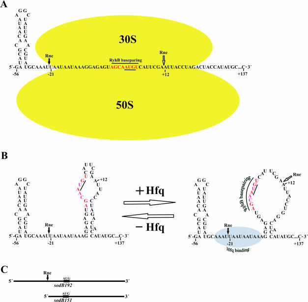
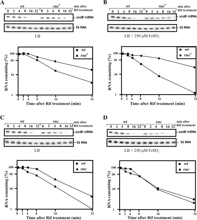
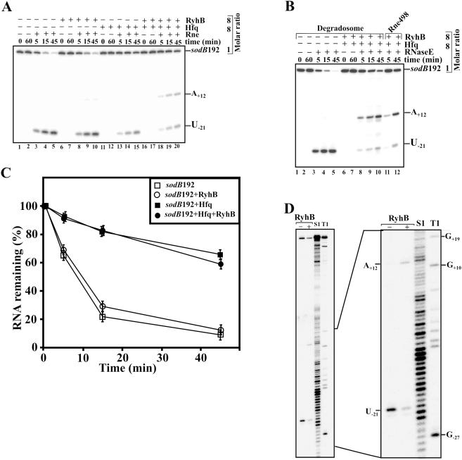
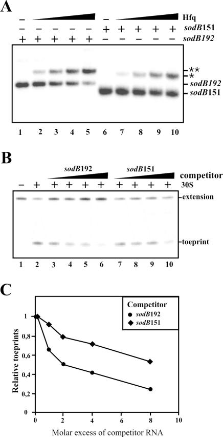
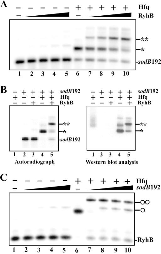
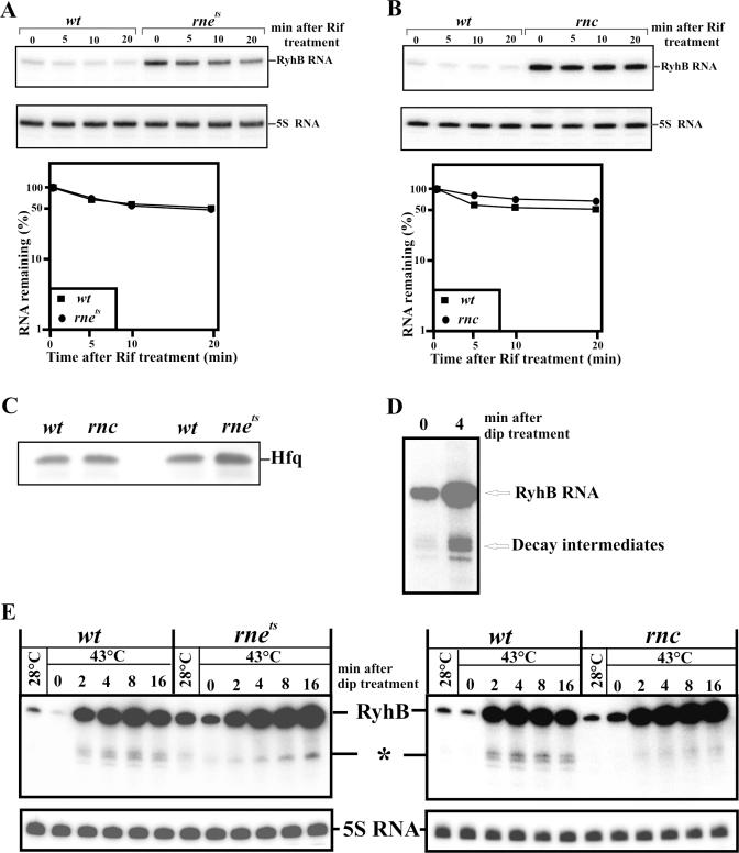
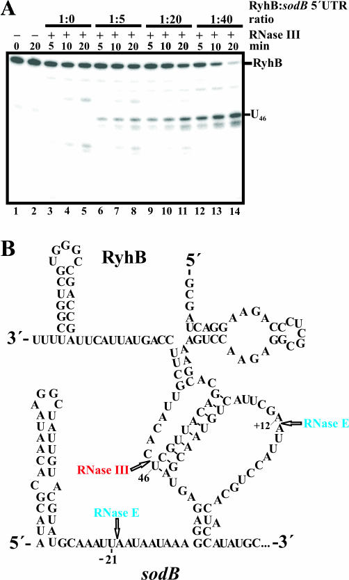
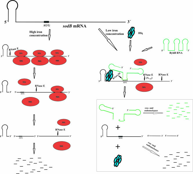
Previous work has demonstrated that iron-dependent variations in the steady-state concentration and translatability of sodB mRNA are modulated by the small regulatory RNA RyhB, the RNA chaperone Hfq and RNase E. In agreement with the proposed role of RNase E, we found that the decay of sodB mRNA is retarded upon inactivation of RNase E in vivo, and that the enzyme cleaves within the sodB 5'-untranslated region (5'-UTR) in vitro, thereby removing the 5' stem-loop structure that facilitates Hfq and ribosome binding. Moreover, RNase E cleavage can also occur at a cryptic site that becomes available upon sodB 5'-UTR/RyhB base pairing. We show that while playing an important role in facilitating the interaction of RyhB with sodB mRNA, Hfq is not tightly retained by the RyhB-sodB mRNA complex and can be released from it through interaction with other RNAs added in trans. Unlike turnover of sodB mRNA, RyhB decay in vivo is mainly dependent on RNase III, and its cleavage by RNase III in vitro is facilitated upon base pairing with the sodB 5'-UTR. These data are discussed in terms of a model, which accounts for the observed roles of RNase E and RNase III in sodB mRNA turnover.


Figures

Figure 1
Figure 1
Alternative structures of the sodB 5′-UTR. (A) 30S ribosome subunit binding and subsequent formation of the translation initiation complex is believed to limit the access of Hfq, RyhB and/or RNase E (Rne) to the 5′ end of the sodB transcript. (B) In the absence of translation, the sodB 5′-leader apparently forms two alternative structures that were previously characterized by Geissman and Touati (28). The transition between these alternative structures is mediated by Hfq and determines the ability of the sodB mRNA to base pair with the small regulatory RNA RyhB (28). Indicated are two regions, which interact with Hfq and base pair with RyhB, respectively. The start codon of the sodB mRNA is underlined. The major RNase E cleavage site (black arrow) and an extra RNase E site (open arrow), which becomes available upon RyhB binding, were mapped during the course of this work (for details, see Figure 3). (C) The bars schematically depict sodB192 and sodB151 mRNAs. The positions of the initiation codon (AUG) and the position of the 5′ terminal RNase E cleavage site are indicated by black boxes and by an arrow, respectively.
Figure 2
Figure 2
RNase E- and RNase III-dependence of sodB mRNA stability in vivo. E.coli strains and isogenic RNase E (rnets) and RNase III (rnc) mutants were grown either in LB medium (LB) or in LB medium supplemented with 250 μM FeSO4 (LB + 250 μM FeSO4) before rifampicin treatment, thereby favouring either RyhB-dependent (A and C) or RyhB-independent (B and D) sodB mRNA decay, respectively. RNA samples prepared from the above cultures before and after rifampicin treatment at the times indicated on top were analysed by northern blotting using probes specific for the sodB transcript and 5S rRNA. The latter was employed as an internal standard for normalization of sodB-specific signals. The graph at the bottom of each panel shows the relative amount of sodB mRNA remaining at each time point as determined by phosphorimaging, and plotted as a function of time.
Figure 3
Figure 3
RNase E cleavage within the sodB 5′-UTR. 5′-[32P]labelled sodB192 was incubated either with RNase E polypeptide (Rne498, residues 1–498) or with RNA degradosome (40) in the presence or absence of Hfq and RyhB at 37°C (A and B, respectively). Aliquots were withdrawn at the times indicated above each lane, phenol extracted and analysed on an 8% sequencing gel. The graph on (C) shows the relative amount of sodB192 mRNA remaining at each time point of (A) as determined by phosphorimaging and plotted as a function of time. (D) Mapping of the RNase E cleavage sites within the sodB 5′-UTR in the presence (+) or absence (−) of RyhB. The molar ratio of Hfq-hexamer:RyhB:sodB192 was 8:8:1, respectively. The precise position of RNase E cleavage sites was determined from concomitantly run S1 and T1 digests of the same RNA.
Figure 4
Figure 4
Effect of the 5′ terminal stem–loop structure on the affinity of the sodB 5′-UTR for Hfq and ribosomes. (A) 5′ end-labelled sodB192 and sodB151 RNAs were incubated alone (lanes 1 and 6) or with increasing amounts (2-, 4-, 6- and 8-fold molar excess) of Hfq-hexamer (lanes 2–5 and 7–10, respectively), and the resulting mixtures were then analysed on a 6% native gel. The positions of free sodB151 and sodB192 RNAs as well as their complexes with Hfq (single and double asterisks, respectively) are indicated. (B) Differential decrease of 30S ribosome binding to sodB192 mRNA by sodB192 and sodB151 competitor RNA, respectively. The sodB192 RNA pre-annealed to the 5′ end-labelled primer was incubated with 30S ribosomal subunits in the presence of increasing amounts (1-, 2-, 4- and 8-fold molar excess) of competitor RNAs (sodB192 and sodB151, respectively). Translation inhibition complex formation was further analysed by primer extension as described in Materials and Methods. (C) Relative toeprints obtained on sodB192 RNA [see (B)] using sodB192 and sodB151 RNA as competitors, respectively. The relative toeprints (%) were calculated as described by Hartz et al. (58) after quantitation of the toeprint and extension signals using the equation: [toeprint signal/(toeprint signal + extension signal)].
Figure 5
Figure 5
Hfq recycling from the RyhB–sodB 5′-UTR complex is facilitated by a molar excess of target RNA. (A) Radioactively labelled sodB192 RNA (nucleotides −56 to 136) was incubated alone (lane 1) or with increasing quantities of RyhB (1-, 2-, 4- and 8-fold molar excess) in the absence (lanes 2–5) or presence (lanes 6–10) of Hfq (the ratio of sodB192 RNA to Hfq-hexamer was 1:1), and the resulting complexes were analysed on a 6% native polyacrylamide gel as described in Materials and Methods. Single and double asterisks indicate the sodB192–Hfq and sodB192–Hfq–RyhB complexes, respectively. (B) Samples containing Hfq alone (lane 1), sodB192 RNA alone (lane 2) or with RyhB (lane 3) as well as its complexes with Hfq in the absence (lane 4) or presence (lane 5) of RyhB were analysed on a 6% native polyacrylamide gel as described in Materials and Methods (left) followed by western blot analysis using anti-Hfq antibodies (right). Single and double asterisks indicate the sodB192–Hfq and sodB192–Hfq–RyhB complexes, respectively. (C) Radioactively labelled RyhB was incubated alone (lane 1), or with increasing quantities of sodB192 RNA (1-, 2-, 4- and 8-fold molar excess; lanes 2–5, respectively) or with the equivalent amount of Hfq in the absence (lane 6) or presence (lane 7) of 1-fold molar excess of sodB192 RNA. Lanes 8–10 correspond to samples containing the pre-formed ternary complex (shown in lane 7), which was further incubated with a 2-, 4- or 8-fold excess of sodB192 RNA, respectively. The resulting complexes were analysed on a 6% native polyacrylamide gel as described in Materials and Methods. Single and double circles indicate the RyhB–sodB192 and RyhB–Hfq–sodB192 complexes, respectively.
Figure 6
Figure 6
Effects of RNase E and RNase III inactivation on RyhB stability in vivo. (A and B) RNA samples prepared from wild-type E.coli cells (wt) and their isogenic RNase E (rne) and RNase III (rnc) mutants at various time points before and after rifampicin treatment were analysed by northern blotting using probes specific for RyhB and 5S rRNA. The latter was employed as an internal standard for normalization of RyhB-specific signals. The graph at the bottom of each panel shows the relative amount of RyhB remaining at each time point as determined by phosphorimaging and plotted as a function of time. (C) Equal amounts of total protein cell extracts prepared from wild-type E.coli cells (wt) and their isogenic RNase E (rne) or RNase III (rnc) mutants were fractionated on a 15% SDS–polyacrylamide gel followed by western blot analysis using anti-Hfq antibodies. The position of Hfq is indicated. (D) RNA samples prepared from wild-type E.coli cells before (0) and after (4) 2,2′-dipyridyl treatment (dip) at 28°C were analysed by northern blotting using a probe specific for RyhB. An asterisk indicates the position of RyhB decay intermediates. (E) RNA samples prepared from wild-type E.coli cells (wt) and their isogenic RNase E (rne) and RNase III (rnc) mutants at various time points before and after 2,2′-dipyridyl treatment (dip) were analysed by northern blotting using probes specific for RyhB and 5S rRNA. The latter was employed as an internal loading control. The molar ratio of Hfq:RyhB was 8:1, respectively. An asterisk indicates the position of RyhB decay intermediates.
Figure 7
Figure 7
RNase III cleavage of RyhB is stimulated by base pairing with its mRNA target. (A) Radioactively labelled RyhB pre-incubated with a 6-fold molar excess of Hfq was further incubated with RNase III in the absence (lanes 1–5) or presence of increasing quantities of sodB192 RNA (5-, 20- and 40-fold molar excess; lanes 6–14, respectively), and aliquots withdrawn at times indicated above each lane were analysed on a 6% (w/v) polyacrylamide sequencing-type gel. The major nucleotide (U46) at which RNase III cleaves RyhB RNA was determined from concomitantly run S1 and T1 digests of the same RNA (data not shown). (B) Model for sodB mRNA–RyhB interaction adopted from Geissmann and Touati (28). The major RNase III site of RyhB [see (A)] and RNase E cleavage sites within the 5′-leader of the sodB transcript (this study) are shown.
Figure 8
Figure 8
Model for sodB mRNA decay at high and low iron concentrations. The pathway for sodB mRNA decay in the presence of steady-state levels of iron is shown on the left. The RNase E cleavage eliminates the 5′ terminal stem–loop structure and triggers both chemical and functional inactivation of the transcript. Owing to lower affinity for 30S ribosomal subunits, translation of the truncated sodB mRNA is less efficient, thereby allowing RNase E cleavage at downstream site(s). The latter results in the subsequent loss of ribosomal subunits and degradation of the intermediate ribosome-free RNA fragments by endo- and exonucleases. The iron-dependent inactivation of the sodB transcript, which is initiated by the small regulatory RNA RyhB and Hfq, is shown on the right. The base pairing with RyhB, which is known to cause structural rearrangements within the sodB 5′-UTR (28), inhibits translation and induces RNase E cleavage at the downstream site A+12, whereas the coordinated decay of RyhB is initiated by RNase III cleavage at U46 (for details, see Figure 7). Similar to the general pathway, the degradation of the intermediate products is accomplished by exo- and endoribonucleases.

References

    1. Le Derout J., Regnier P., Hajnsdorf E. Both temperature and medium composition regulate RNase E processing efficiency of the rpsO mRNA coding for ribosomal protein S15 of Escherichia coli. J. Mol. Biol. 2002;319:341–349. - PubMed
    1. Nilsson G., Belasco J.G., Cohen S.N., von Gabain A. Growth-rate dependent regulation of mRNA stability in Escherichia coli. Nature. 1984;312:75–77. - PubMed
    1. Vytvytska O., Jakobsen J.S., Balcunaite G., Andersen J.S., Baccarini M., von Gabain A. Host factor I, Hfq, binds to Escherichia coli ompA mRNA in a growth rate dependent fashion and regulates its stability. Proc. Natl Acad. Sci. USA. 1998;95:14118–14123. - PMC - PubMed
    1. Coburn G.A., Mackie G.A. Degradation of mRNA in Escherichia coli: an old problem with some new twists. Prog. Nucleic Acid Res. Mol. Biol. 1999;62:55–108. - PubMed
    1. Lundberg U., Kaberdin V., von Gabain A. The mechanisms of mRNA degradation in bacteria and their implication for stabilization of heterologous transcripts. In: Demain A.L., Davies R.M., Cohen G., Hershberg C.L., Sherman D.H., Willson R.C., Wu J.-H.D., editors. Manual of Industrial Microbiology and Biotechnology. 2nd edn. Washington, DC: ASM Press; 1999. pp. 585–596.

Publication types

MeSH terms