. 2005 Mar 21;201(6):871-9.
doi: 10.1084/jem.20041497.

Platelet activation leads to activation and propagation of the complement system

Affiliations

Platelet activation leads to activation and propagation of the complement system

Ian Del Conde et al. J Exp Med. .

Abstract

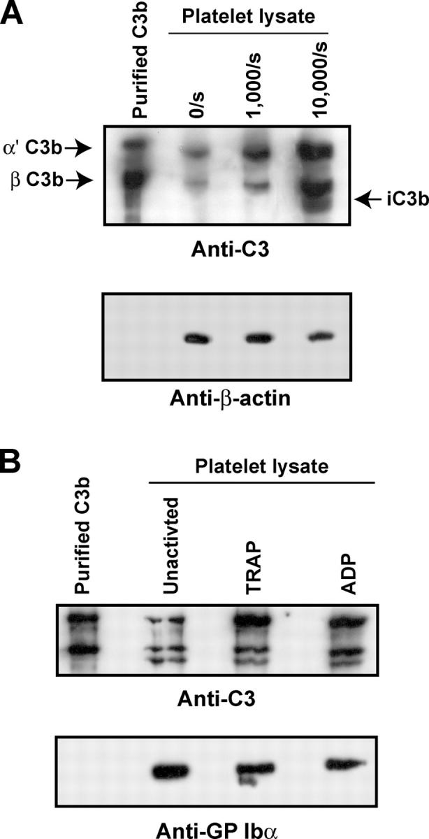
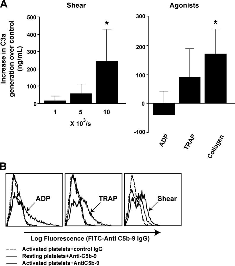
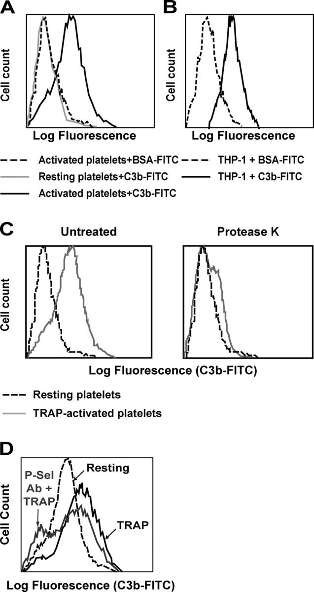
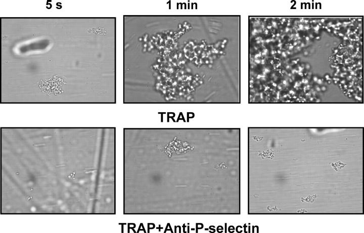
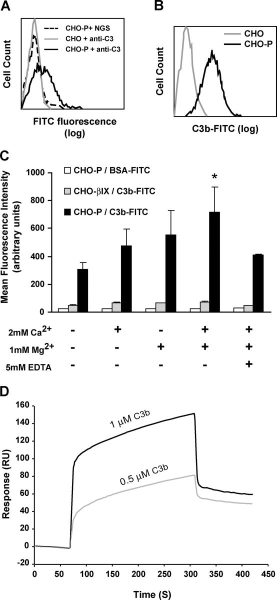
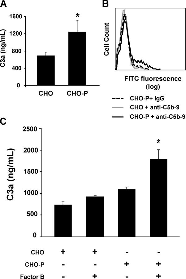
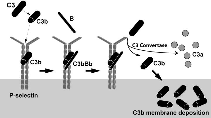
Inflammation and thrombosis are two responses that are linked through a number of mechanisms, one of them being the complement system. Various proteins of the complement system interact specifically with platelets, which, in turn, activates them and promotes thrombosis. In this paper, we show that the converse is also true: activated platelets can activate the complement system. As assessed by flow cytometry and immunoblotting, C3 deposition increased on the platelet surface upon cell activation with different agonists. Activation of the complement system proceeded to its final stages, which was marked by the increased generation of the anaphylotoxin C3a and the C5b-9 complex. We identified P-selectin as a C3b-binding protein, and confirmed by surface plasmon resonance binding that these two proteins interact specifically with a dissociation constant of 1 microM. Using heterologous cells expressing P-selectin, we found that P-selectin alone is sufficient to activate the complement system, marked by increases in C3b deposition, C3a generation, and C5b-9 formation. In summary, we have found that platelets are capable of activating the complement system, and have identified P-selectin as a receptor for C3b capable of initiating complement activation. These findings point out an additional mechanism by which inflammation may localize to sites of vascular injury and thrombosis.


Figures

Figure 1.
Figure 1.
Deposition of C3 on activated platelets. Platelet-rich plasma was (A) subjected to different shear rates for 60 s, or (B) activated with different agonists. Platelets were then pelleted, washed with TBS buffer, lysed in reducing SDS sample buffer, and subjected to PAGE and Western blotting for C3. The same membranes was stripped and probed for β-actin or GP Ibα as loading controls. These results are representative of three experiments.
Figure 2.
Figure 2.
Activation of complement on activated platelets. (A) Generation of C3a by activated platelets. 1 ml PRP was stimulated with either 20 μM ADP, 10 μM TRAP, 20 μg/ml collagen, or with different shear rates for 60 s. Platelets were pelleted, and C3a levels were measured in the plasma supernatant using ELISA. The plasma of a suspension of resting platelets served as the controls. n = 4; *, P < 0.05. (B) Formation of the C5b-9 complex on activated platelets: platelet-rich plasma (PRP) was stimulated with either 20 μM ADP, 10 μM TRAP, or 10,000 s−1 × 60-s shear. Platelets were then probed by flow cytometry for the presence of the C5b-9 complex using a rabbit anti–C5b-9 neoepitope. Baseline fluorescence was set using nonimmune rabbit IgG and a secondary FITC-conjugated antibody. These results are representative of four experiments.
Figure 3.
Figure 3.
Binding of C3b to activated platelets. (A) Binding of C3b to the surface of activated platelets was measured by flow cytometry using FITC-conjugated purified C3b. Washed platelets, either resting or stimulated with 5 μM TRAP, were probed with FITC-C3b. Background fluorescence was set using BSA-FITC. (B) Binding of C3b-FITC to the monocytic cells, THP-1, which constitutively express the complement receptor CR1. (C) Cleavage of surface proteins inactivated using 50 μg/ml protease K abrogates the binding of FITC-conjugated C3b. (D) Binding of FITC-C3b to TRAP-activated platelets was inhibited by treating the platelets with anti–P-selectin antibody.
Figure 4.
Figure 4.
P-Selectin mediates the interaction between activated platelets and C3b. TRAP-activated platelets were perfused over C3b immobilized on a glass coverslip (30 μg/ml) at a constant shear stress of 2 dyne/cm2 in a parallel plate flow chamber. Thrombus formation was visualized and recorded over 2 min. Treatment of platelets with an anti– P-selectin polyclonal antibody (25 μg/ml) abolished platelet adhesion to the immobilized C3b. Activated platelets failed to adhere to immobilized BSA. All the images are taken with 20× magnification.
Figure 5.
Figure 5.
Binding of C3b to P-selectin. (A) Binding of plasma C3 to CHO-P and CHO cells was studied by incubating the cells in platelet-poor plasma for 30 min at 37°C. Cells were then probed with goat anti-C3 antiserum and a secondary FITC-anti–goat antibody. Baseline fluorescence was set using normal goat serum (NGS) and the secondary antibody. (B) Binding of FITC-labeled purified C3b was determined in CHO and CHO-P by flow cytometry. (C) The effect of calcium and magnesium on the binding of C3b to P-selectin expressed on CHO cells (CHO-P) was assessed by flow cytometry. Binding of FITC-labeled bovine serum albumin (BSA-FITC) to P-selectin, and binding of C3b-FITC to cells expressing the βIX subunit of the GPIb–IX-V complex served as negative controls. n = 4; *, P < 0.05. (D) Surface plasmon resonance binding studies of the interaction between purified C3b and recombinant ectodomain of P-selectin. Binding curves of P-selectin were obtained perfusing different concentrations of purified C3b over the P-selectin surface, as described in the Materials and methods. Dissociation constant (K d) obtained for binding of C3b to P-selectin was 1.0 μM. RU, resonance units.
Figure 6.
Figure 6.
P-Selectin activates the complement system. P-Selectin—mediated activation of the complement system was studied in CHO cells. (A) CHO-P and CHO cells were incubated in PPP for 60 min. The cells were then sedimented, and C3a was measured in the plasma supernatant by ELISA. n = 4; *, P < 0.05. (B) Formation of the C5b-9 complex on CHO-P and CHO cells was measured by flow cytometry using a rabbit anti–C5b-9 neoepitope. Baseline fluorescence was set using nonimmune rabbit IgG. These results are representative of four other experiments. (C) CHO and CHO-P cells were incubated in a reconstituted complement system, with or without factor B, for 60 min at 37°C. Cells were then sedimented and C3a was measured by ELISA in the supernatants. n = 4; *, P < 0.05.
Figure 7.
Figure 7.
The proposed model P-selectin captures and immobilizes labile C3b. Binding of factor B to C3b initiates formation of C3 convertase (C3bBb) on the surface of activated platelet or endothelial cells. The C3 convertase complex cleaves C3 and mediates amplification of the complement activation (see Discussion for details).

References

    1. Mastellos, D., and J.D. Lambris. 2002. Complement: more than a “guard” against invading pathogens? Trends Immunol. 23:485–491. - PubMed
    1. Gralnick, H.R., M. Vail, L.P. McKeown, P. Merryman, O. Wilson, I. Chu, and J. Kimball. 1995. Activated platelets in paroxysmal nocturnal haemoglobinuria. Br. J. Haematol. 91:697–702. - PubMed
    1. Qin, X., N. Krumrei, L. Grubissich, M. Dobarro, H. Aktas, G. Perez, and J.A. Halperin. 2003. Deficiency of the mouse complement regulatory protein mCd59b results in spontaneous hemolytic anemia with platelet activation and progressive male infertility. Immunity. 18:217–227. - PubMed
    1. Polley, M.J., and R. Nachman. 1978. The human complement system in thrombin-mediated platelet function. J. Exp. Med. 147:1713–1726. - PMC - PubMed
    1. Polley, M.J., and R.L. Nachman. 1983. Human platelet activation by C3a and C3a des-arg. J. Exp. Med. 158:603–615. - PMC - PubMed

Publication types