. 2005 Mar 21;201(6):981-91.
doi: 10.1084/jem.20042311.

In vivo depletion of lung CD11c+ dendritic cells during allergen challenge abrogates the characteristic features of asthma

Affiliations

In vivo depletion of lung CD11c+ dendritic cells during allergen challenge abrogates the characteristic features of asthma

Leonie S van Rijt et al. J Exp Med. .

Abstract

Although dendritic cells (DCs) play an important role in sensitization to inhaled allergens, their function in ongoing T helper (Th)2 cell-mediated eosinophilic airway inflammation underlying bronchial asthma is currently unknown. Here, we show in an ovalbumin (OVA)-driven murine asthma model that airway DCs acquire a mature phenotype and interact with CD4(+) T cells within sites of peribronchial and perivascular inflammation. To study whether DCs contributed to inflammation, we depleted DCs from the airways of CD11c-diphtheria toxin (DT) receptor transgenic mice during the OVA aerosol challenge. Airway administration of DT depleted CD11c(+) DCs and alveolar macrophages and abolished the characteristic features of asthma, including eosinophilic inflammation, goblet cell hyperplasia, and bronchial hyperreactivity. In the absence of CD11c(+) cells, endogenous or adoptively transferred CD4(+) Th2 cells did not produce interleukin (IL)-4, IL-5, and IL-13 in response to OVA aerosol. In CD11c-depleted mice, eosinophilic inflammation and Th2 cytokine secretion were restored by adoptive transfer of CD11c(+) DCs, but not alveolar macrophages. These findings identify lung DCs as key proinflammatory cells that are necessary and sufficient for Th2 cell stimulation during ongoing airway inflammation.


Figures

Figure 1.
Figure 1.
Phenotype of DCs in bronchoalveolar compartment, lung, and lung draining LNs in steady-state and during inflammation. (A) BALB/c mice were OVA sensitized and challenged (OVA/OVA), or as a control sham sensitized/challenged (PBS/PBS), to induce an eosinophilic airway inflammation. DCs were analyzed for the expression of molecules needed for T cell interaction. DCs were recognized as CD11c+/MHCII+ (top). Black lines indicate marker expression and gray lines indicate the isotype control staining. (B) 1 d after the last aerosol, peribronchial infiltrates were analyzed for the presence of CD4+ T cells (blue) and CD11c+ DCs (red) in frozen lung sections. Bar, 100 μm. (C) At a larger magnification, it is shown that DCs and T cells are interacting in the peribronchial infiltrates as well as in the lung parenchyma. Bar, 10 μm.
Figure 2.
Figure 2.
DT treatment depletes CD11c+ cells in CD11c-DTR Tg mice. Naive CD11c-DTR Tg and nTg mice were i.t. injected with 50 ng DT (Tg DT and nTG DT) or only PBS (Tg PBS). (A, left) 48 h after instillation, BALF cells, lungs, and lung draining LNs were analyzed for the presence of CD11c+MHCII+ DCs by FACS. (right) Systemic effects of DT on peripheral axillary LNs and splenic DCs. (B) OVA-sensitized Tg and nTg mice were treated with DT or PBS and received three OVA aerosols. Lung sections stained for CD4+ (blue) and CD11c+ (red) cells showed that i.t. administration of 50 ng DT in Tg mice before challenge, inhibited the induction of peribronchial infiltrates containing CD4+ and CD11c+ cells (left), whereas in nTg mice (right) DT did not affect CD11c+ DCs. As a control, Tg mice were treated with PBS (middle), showing a similar peribronchial infiltrate as in WT mice. The results shown represent two independent experiments. Bars, 100 μm.
Figure 3.
Figure 3.
DC requirement for induction of secondary immune responses. Mice were OVA sensitized at day 0, received a DT i.t. injection at day 10, and received a daily 30-min aerosol at days 11–13. At day 14, mice were killed. (A) BALF cells were analyzed for total BALF numbers and (B) the number of eosinophils 1 d after the last aerosol. (C) AHR was measured in CD11c-DTR Tg mice and nTg littermates that were sensitized with OVA (alum) and challenged with three OVA aerosols or as a control with PBS aerosols. 24 h before the first OVA aerosol, all mice were treated with DT. Tg DC/OVA, OVA-challenged DC-depleted CD11c-DTR Tg; nTg DC+/OVA, OVA-challenged nTg mice; Tg DC/PBS, PBS-challenged DC-depleted CD11c-DTR Tg; nTg DC+/PBS, PBS-challenged nTg. (D) Th2 cytokine production by lung draining LNs in Tg DC, Tg DC+, and nTg DC+ mice. (E) A cohort of in vitro–obtained memory Th2 were adoptively transferred (i.v.) into Tg DC and nTg DC+, simultaneously with an i.t. injection of DT or as a control PBS (Tg DC+). 24 h after the adoptive transfer, mice were challenged with OVA aerosols for 4 d. Lung draining LNs were restimulated ex vivo with OVA and IL-4, IL-5, IL-13, and IFNγ levels were determined in supernatant by ELISA (*, P < 0.05 vs. nTg DC+). The results shown represent one out of five independent experiments and are expressed as means ± SEM.
Figure 4.
Figure 4.
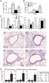
DC reconstitution restored eosinophilic airway inflammation. CD11c-DTR Tg and nTg mice were OVA sensitized at day 0, received i.t. DT (Tg DC, Tg DC + DC i.t. and nTg DC+) or PBS (Tg DC+) at day 10 and were challenged with OVA aerosols at days 11–13. Mice were reconstituted with unpulsed 2 × 10⁶ DCs i.t. immediately after OVA challenge. BALF was analyzed for (A) total BALF numbers and (B) the number of eosinophils at day 14. (C) Th2 cytokine production by MLN after ex vivo restimulation with OVA was measured in supernatant by ELISA (*, P < 0.05 vs. Tg DC). (D–G) Histological sections of Tg DC, Tg DC + DC i.t., Tg DC+, and nTg DC+ mice stained with PAS reagent. Bars, 100 μm. (H) Number of eosinophils in BALF after reconstitution with macrophages, CD8+ cells or eosinophils (*, P < 0.05 vs. C). The results shown represent one out of two independent experiments and are expressed as means ± SEM.
Figure 5.
Figure 5.
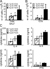
DC requirement for maintaining eosinophilic airway inflammation. CD11c-DTR Tg (Tg DC/OVA) and nTg mice (nTg DC+/OVA) were OVA sensitized at day 0, received a daily 30-min aerosol at days 11–13. At day 14, mice received DT, and at days 15–17 mice were challenged daily with a 30-min OVA. One group of CD11c-DTR Tg mice (Tg DC/PBS) received the same treatment but was challenged with PBS aerosol after DC depletion. BALF was analyzed for (A) total BALF numbers and (B) the number of eosinophils. (C) Th2 cytokine production by MLN after ex vivo restimulation with OVA was measured in supernatant by ELISA. (D–F) Histological sections of Tg DC/OVA, Tg DC/PBS, and nTg DC+/OVA mice stained with PAS reagent (*, P < 0.05 vs. nTg DC+ /OVA). The results shown represent one out of three independent experiments and are expressed as means ± SEM. Bars, 100 μm.
Figure 5.
Figure 5.
DC requirement for maintaining eosinophilic airway inflammation. CD11c-DTR Tg (Tg DC/OVA) and nTg mice (nTg DC+/OVA) were OVA sensitized at day 0, received a daily 30-min aerosol at days 11–13. At day 14, mice received DT, and at days 15–17 mice were challenged daily with a 30-min OVA. One group of CD11c-DTR Tg mice (Tg DC/PBS) received the same treatment but was challenged with PBS aerosol after DC depletion. BALF was analyzed for (A) total BALF numbers and (B) the number of eosinophils. (C) Th2 cytokine production by MLN after ex vivo restimulation with OVA was measured in supernatant by ELISA. (D–F) Histological sections of Tg DC/OVA, Tg DC/PBS, and nTg DC+/OVA mice stained with PAS reagent (*, P < 0.05 vs. nTg DC+ /OVA). The results shown represent one out of three independent experiments and are expressed as means ± SEM. Bars, 100 μm.
Figure 6.
Figure 6.
Adoptive transfer of DCs is sufficient to induce all asthmatic features in sensitized mice. Mice were OVA DC sensitized at day 0, received at days 10 and 12 an i.t. injection of OVA DCs (black bars), or received at days 10–13 a daily OVA aerosol (white bars) or PBS aerosols (diagonally striped bars). (A and B) At day 14, total BALF cells numbers and eosinophil numbers were determined by FACS (*, P < 0.05 vs. DC/PBS). (C) Th2 cytokine production by MLNs was determined after ex vivo restimulation with OVA. IL-4, IL-5, IL-13, and IFN-γ were measured in supernatant by ELISA. (D–F) Histological lung sections of mice challenged with OVA DCs (DC/DC), OVA aerosols (DC/OVA), and PBS aerosols (DC/PBS). (G) Airway responsiveness was increased in DC/DC and DC/OVA compared with DC/PBS mice. The results represent one out of two independent experiments and are expressed as means ± SEM.
Figure 6.
Figure 6.
Adoptive transfer of DCs is sufficient to induce all asthmatic features in sensitized mice. Mice were OVA DC sensitized at day 0, received at days 10 and 12 an i.t. injection of OVA DCs (black bars), or received at days 10–13 a daily OVA aerosol (white bars) or PBS aerosols (diagonally striped bars). (A and B) At day 14, total BALF cells numbers and eosinophil numbers were determined by FACS (*, P < 0.05 vs. DC/PBS). (C) Th2 cytokine production by MLNs was determined after ex vivo restimulation with OVA. IL-4, IL-5, IL-13, and IFN-γ were measured in supernatant by ELISA. (D–F) Histological lung sections of mice challenged with OVA DCs (DC/DC), OVA aerosols (DC/OVA), and PBS aerosols (DC/PBS). (G) Airway responsiveness was increased in DC/DC and DC/OVA compared with DC/PBS mice. The results represent one out of two independent experiments and are expressed as means ± SEM.

References

    1. Wills-Karp, M. 1999. Immunologic basis of antigen-induced airway hyperresponsiveness. Annu. Rev. Immunol. 17:255–281. - PubMed
    1. Herrick, C.A., and K. Bottomly. 2003. To respond or not to respond: T cells in allergic asthma. Nat. Rev. Immunol. 3:405–412. - PubMed
    1. Holt, P.G., M.A. Schon-Hegrad, and J. Oliver. 1988. MHC class II antigen-bearing dendritic cells in pulmonary tissues of the rat. Regulation of antigen presentation activity by endogenous macrophage populations. J. Exp. Med. 167:262–274. - PMC - PubMed
    1. Banchereau, J., and R.M. Steinman. 1998. Dendritic cells and the control of immunity. Nature. 392:245–252. - PubMed
    1. Brimnes, M.K., L. Bonifaz, R.M. Steinman, and T.M. Moran. 2003. Influenza virus-induced dendritic cell maturation is associated with the induction of strong T cell immunity to a coadministered, normally nonimmunogenic protein. J. Exp. Med. 198:133–144. - PMC - PubMed

Publication types

MeSH terms