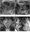
Assessment of the prognostic factors for a local recurrence of rectal cancer: the utility of preoperative MR imaging
- PMID: 15782014
- PMCID: PMC2685000
- DOI: 10.3348/kjr.2005.6.1.8
Assessment of the prognostic factors for a local recurrence of rectal cancer: the utility of preoperative MR imaging
Abstract
Objective: To determine the utility of MR imaging in evaluating the prognostic factors for a local recurrence of rectal cancer following a curative resection.
Materials and methods: The preoperative MR images obtained from 17 patients with a local recurrence and 54 patients without a local recurrence, who had undergone a curative resection, were independently evaluated by three radiologists. The following findings were analyzed: the direct invasion of the perirectal fat by the primary rectal carcinoma, involvement of the perirectal lymph nodes, perirectal spiculate nodules, perivascular encasement, and an enlargement of the pelvic wall lymph nodes. The clinical and surgical profiles were obtained from the patients' medical records. The association of a local recurrence with the MR findings and the clinicosurgical variables was statistically evaluated.
Results: Of the MR findings, the presence of perivascular encasement (p = 0.001) and perirectal spiculate nodules (p = 0.001) were found to be significant prognostic factors for a local recurrence. Of the clinicosurgical profiles, the presence of a microscopic vascular invasion (p = 0.005) and the involvement of the regional lymph nodes (p = 0.006) were associated with a local recurrence. Logistic regression analysis showed that the presence of perirectal spiculate nodules was an independent predictor of a local recurrence (odds ratio, 7.382; 95% confidence interval, 1.438, 37.889; p = 0.017).
Conclusion: The presence of perirectal spiculate nodules and perivascular encasement on the preoperative MR images are significant predictors of a local recurrence after curative surgery for a rectal carcinoma. This suggests that preoperative MR imaging can provide useful information to help in the planning of preoperative adjuvant therapy.
Figures


References
-
- Ross A, Rusnak C, Weinerman B, Kuechler P, Hayashi A, Maclachlan G, et al. Recurrence and survival after surgical management of rectal cancer. Am J Surg. 1999;177:392–395. - PubMed
-
- Tveit KM, Guldvog I, Hagen S, Trondsen E, Harbitz T, Nygaard K, et al. Norwegian Adjuvant Rectal Cancer Project Group. Randomized controlled trial of postoperative radiotherapy and short-term time-scheduled 5-fluorouracil against surgery alone in the treatment of Dukes B and C rectal cancer. Br J Surg. 1997;84:1130–1135. - PubMed
-
- Heald RJ, Ryall RD. Recurrence and survival after total mesorectal excision for rectal cancer. Lancet. 1986;1:1479–1482. - PubMed
-
- Reynolds JV, Joyce WP, Dolan J, Sheahan K, Hyland JM. Pathological evidence in support of total mesorectal excision in the management of rectal cancer. Br J Surg. 1996;83:1112–1115. - PubMed
-
- Kapiteijn E, Marijnen CA, Nagtegaal ID, Putter H, Steup WH, Wiggers T, et al. Preoperative radiotherapy combined with total mesorectal excision for resectable rectal cancer. N Engl J Med. 2001;345:638–646. - PubMed
MeSH terms
LinkOut - more resources
Full Text Sources
Medical

