

CT and MR imaging of the buccal space: normal anatomy and abnormalities
- PMID: 15782016
- PMCID: PMC2684993
- DOI: 10.3348/kjr.2005.6.1.22
CT and MR imaging of the buccal space: normal anatomy and abnormalities
Abstract
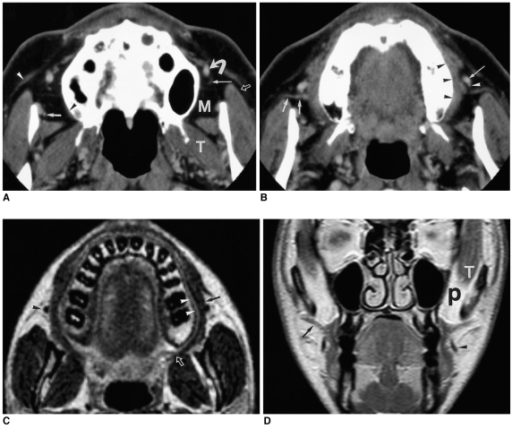
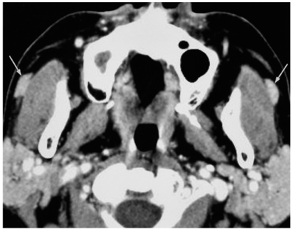
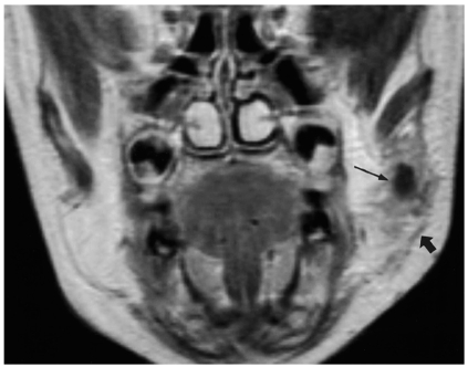
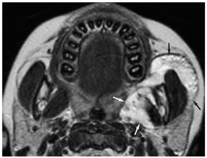
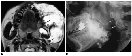
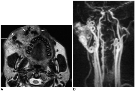
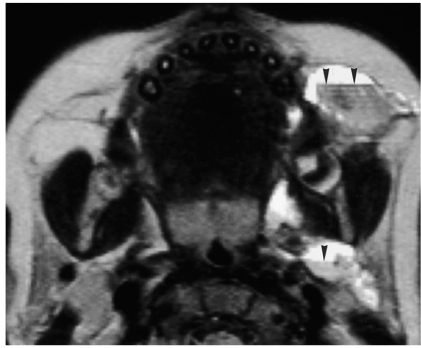
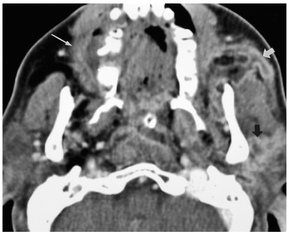
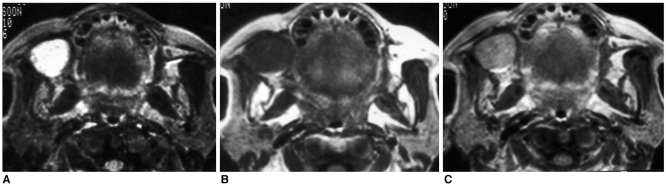
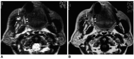
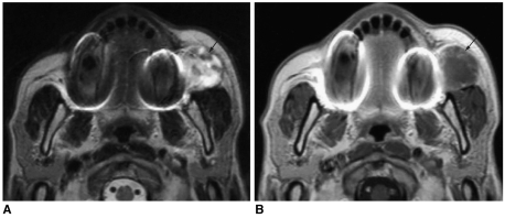
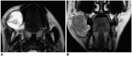
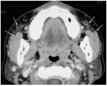
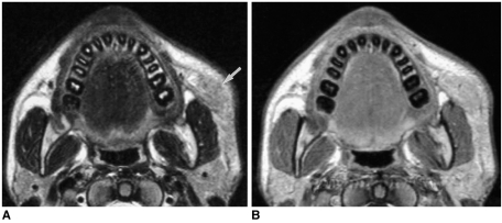
The buccal space is an anatomical compartment lying anterior to the masticator space and lateral to the buccinator muscle. Since the major purpose of imaging is to define the likely anatomic origin and also the extent of a given lesion, thorough knowledge of the normal anatomy of the buccal space is essential, and this knowledge can aid the physician in narrowing down the list of possible maladies on the differential diagnosis. We illustrate here in this paper the important anatomic landmarks and typical pathologic conditions of the buccal space such as the developmental lesions and the neoplastic lesions. Knowledge of the expected pathologic conditions is useful for the radiologist when interpreting facial CT and MR images.
Figures
References
-
- Tart RP, Kotzur IM, Mancuso AA, Glantz MS, Mukherji SK. CT and MR imaging of the buccal space and buccal space masses. RadioGraphics. 1995;15:531–550. - PubMed
-
- Smoker WRK. Oral cavity. In: Som PM, Curtin HD, editors. Head and neck imaging. 3rd ed. St. Louis: Mosby; 1996. pp. 488–544.
-
- Kurabayashi T, Ida M, Tetsumura A, Ohbayashi N, Yasumoto M, Sasaki T. MR imaging of benign and malignant lesions in the buccal space. Dentomaxillofac Radiol. 2002;31:344–349. - PubMed
-
- Kurabayashi T, Ida M, Yoshino N, Sasaki T, Kishi T, Kusama M. Computed tomography in the diagnosis of buccal space masses. Dentomaxillofac Radiol. 1997;26:347–353. - PubMed
-
- Werner JA, Dunne AA, Folz BJ, Rochels R, Bien S, Ramaswamy A, et al. Current concepts in the classification, diagnosis and treatment of hemangiomas and vascular malformations of the head and neck. Eur Arch Otorhinolaryngol. 2001;258:141–149. - PubMed
