

Ductographic findings of breast cancer
- PMID: 15782017
- PMCID: PMC2684994
- DOI: 10.3348/kjr.2005.6.1.31
Ductographic findings of breast cancer
Abstract
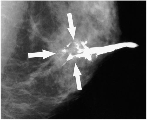
Ductography has become the gold standard for the evaluation of patients exhibiting pathologic nipple discharges. In nine patients (age range, 29-67 years; median age, 51 years) with invasive (n=5) or intraductal (n=4) cancer, ductographic findings were recorded, then correlated with mammographic and sonographic findings. Common ductographic findings included complete ductal obstruction, multiple irregular filling defects in the nondilated peripheral ducts, ductal wall irregularities, periductal contrast extravasation, and ductal displacement. Faint microcalcifications or ill-defined masses, which were not opacified by contrast material, were often discovered adjacent to ductal abnormalities. Mammographically and sonographically occult diffusely spreading intraductal cancers often manifested as pathologic nipple discharge. In such cases, meticulous ductographic examinations and interpretations were crucial in order not to miss breast cancers.
Figures


References
-
- Tabar L, Dean PB, Pentek Z. Galactography: the diagnostic procedure of choice for nipple discharge. Radiology. 1983;149:31–38. - PubMed
-
- Sickles EA. Galactography and other imaging investigations of nipple discharge. Lancet. 2000;356:1622–1623. - PubMed
-
- Slawson SH, Johnson BA. Ductography: how to and what if? RadioGraphics. 2001;21:133–150. - PubMed
-
- Stavros AT. Nontargeted indications: breast secretions, nipple discharge, and intraductal papillary lesions of the breast. In: Stavros AT, editor. Breast ultrasound. 1st ed. Philadelphia, Pa: Lippincott Williams & Wilkins; 2004. pp. 157–198.
-
- Moon WK, Myung JS, Lee YJ, Park IA, Noh DY, Im JG. US of ductal carcinoma in situ. RadioGraphics. 2002;22:269–280. - PubMed
MeSH terms
LinkOut - more resources
Full Text Sources
Medical
