

Rhodococcus equi-specific cytotoxic T lymphocytes in immune horses and development in asymptomatic foals
- PMID: 15784549
- PMCID: PMC1087435
- DOI: 10.1128/IAI.73.4.2083-2093.2005
Rhodococcus equi-specific cytotoxic T lymphocytes in immune horses and development in asymptomatic foals
Abstract
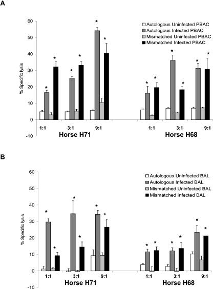
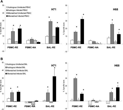
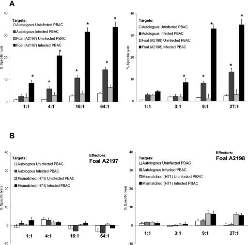
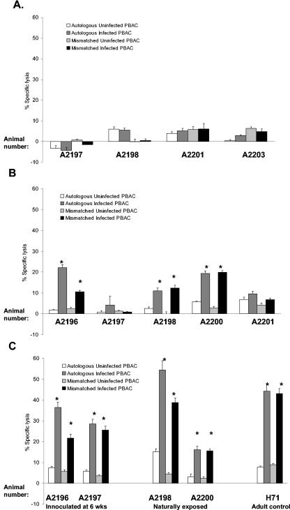
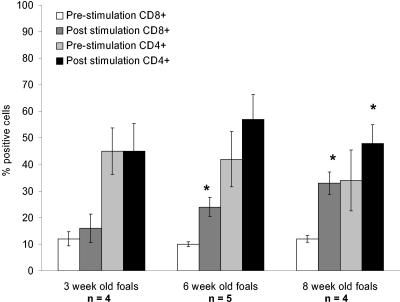
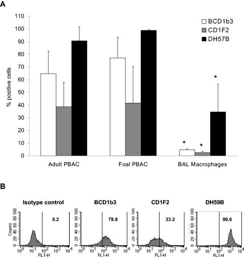
Rhodococcus equi is an important cause of pneumonia in young horses; however, adult horses are immune due to their ability to mount protective recall responses. In this study, the hypothesis that R. equi-specific cytotoxic T lymphocytes (CTL) are present in the lung of immune horses was tested. Bronchoalveolar lavage (BAL)-derived pulmonary T lymphocytes stimulated with R. equi lysed infected alveolar macrophages and peripheral blood adherent cells (PBAC). As with CTL obtained from the blood, killing of R. equi-infected targets by pulmonary effectors was not restricted by equine lymphocyte alloantigen-A (ELA-A; classical major histocompatibility complex class I), suggesting a novel or nonclassical method of antigen presentation. To determine whether or not CTL activity coincided with the age-associated susceptibility to rhodococcal pneumonia, CTL were evaluated in foals. R. equi-stimulated peripheral blood mononuclear cells (PBMC) from 3-week-old foals were unable to lyse either autologous perinatal or mismatched adult PBAC targets. The defect was not with the perinatal targets, as adult CTL effectors efficiently killed infected targets from 3-week-old foals. In contrast, significant CTL activity was present in three of five foals at 6 weeks of age, and significant specific lysis was induced by PBMC from all foals at 8 weeks of age. As with adults, lysis was ELA-A unrestricted. Two previously described monoclonal antibodies, BCD1b3 and CD1F2/1B12.1, were used to examine the expression of CD1, a nonclassical antigen-presenting molecule, on CTL targets. These antibodies cross-reacted with both foal and adult PBAC. However, neither antibody bound alveolar macrophages, suggesting that the R. equi-specific, major histocompatibility complex-unrestricted lysis is not restricted by a surface molecule identified by these antibodies.
Figures
Similar articles
-
The influence of age and Rhodococcus equi infection on CD1 expression by equine antigen presenting cells.Vet Immunol Immunopathol. 2009 Aug 15;130(3-4):197-209. doi: 10.1016/j.vetimm.2009.02.007. Epub 2009 Feb 14. Vet Immunol Immunopathol. 2009. PMID: 19285733
-
Hematologic and immunophenotypic factors associated with development of Rhodococcus equi pneumonia of foals at equine breeding farms with endemic infection.Vet Immunol Immunopathol. 2004 Jul;100(1-2):33-48. doi: 10.1016/j.vetimm.2004.02.010. Vet Immunol Immunopathol. 2004. PMID: 15182994
-
Rhodococcus equi-infected macrophages are recognized and killed by CD8+ T lymphocytes in a major histocompatibility complex class I-unrestricted fashion.Infect Immun. 2004 Dec;72(12):7073-83. doi: 10.1128/IAI.72.12.7073-7083.2004. Infect Immun. 2004. PMID: 15557631 Free PMC article.
-
Current understanding of the equine immune response to Rhodococcus equi. An immunological review of R. equi pneumonia.Vet Immunol Immunopathol. 2010 May 15;135(1-2):1-11. doi: 10.1016/j.vetimm.2009.12.004. Epub 2009 Dec 23. Vet Immunol Immunopathol. 2010. PMID: 20064668 Review.
-
Rhodococcus equi pneumonia in the foal--part 1: pathogenesis and epidemiology.Vet J. 2012 Apr;192(1):20-6. doi: 10.1016/j.tvjl.2011.08.014. Epub 2011 Oct 19. Vet J. 2012. PMID: 22015138 Review.
Cited by
-
IgG Subtype Response against Virulence-Associated Protein A in Foals Naturally Infected with Rhodococcus equi.Vet Sci. 2024 Sep 9;11(9):422. doi: 10.3390/vetsci11090422. Vet Sci. 2024. PMID: 39330801 Free PMC article.
-
Equine neonates have attenuated humoral and cell-mediated immune responses to a killed adjuvanted vaccine compared to adult horses.Clin Vaccine Immunol. 2010 Dec;17(12):1896-902. doi: 10.1128/CVI.00328-10. Epub 2010 Oct 13. Clin Vaccine Immunol. 2010. PMID: 20943883 Free PMC article.
-
Early development of cytotoxic T lymphocytes in neonatal foals following oral inoculation with Rhodococcus equi.Vet Immunol Immunopathol. 2011 Jun 15;141(3-4):312-6. doi: 10.1016/j.vetimm.2011.03.015. Epub 2011 Mar 21. Vet Immunol Immunopathol. 2011. PMID: 21481947 Free PMC article.
-
Protective immune response against Rhodococcus equi: An innate immunity-focused review.Equine Vet J. 2025 May;57(3):563-586. doi: 10.1111/evj.14214. Epub 2024 Sep 11. Equine Vet J. 2025. PMID: 39258739 Free PMC article. Review.
-
Evaluation of vaccine candidates against Rhodococcus equi in BALB/c mice infection model: cellular and humoral immune responses.BMC Microbiol. 2024 Jul 8;24(1):249. doi: 10.1186/s12866-024-03408-z. BMC Microbiol. 2024. PMID: 38977999 Free PMC article.
References
-
- Adkins, B. 2000. Development of neonatal Th1/Th2 function. Int. Rev. Immunol. 19:157-171. - PubMed
-
- Adkins, B. 1999. T-cell function in newborn mice and humans. Immunol. Today 20:330-335. - PubMed
-
- Adkins, B., and R. Q. Du. 1998. Newborn mice develop balanced Th1/Th2 primary effector responses in vivo but are biased to Th2 secondary responses. J. Immunol. 160:4217-4224. - PubMed
-
- Alvarez, B., C. Sanchez, R. Bullido, A. Marina, J. Lunney, F. Alonso, A. Ezquerra, and J. Dominguez. 2000. A porcine cell surface receptor identified by monoclonal antibodies to SWC3 is a member of the signal regulatory protein family and associates with protein-tyrosine phosphatase SHP-1. Tissue Antigens 55:342-351. - PubMed
Publication types
MeSH terms
Substances
Grants and funding
LinkOut - more resources
Full Text Sources
Research Materials
