. 2005 Apr;73(4):2101-8.
doi: 10.1128/IAI.73.4.2101-2108.2005.

Interleukin 10- and Fcgamma receptor-deficient mice resolve Leishmania mexicana lesions

Affiliations

Interleukin 10- and Fcgamma receptor-deficient mice resolve Leishmania mexicana lesions

Laurence U Buxbaum et al. Infect Immun. 2005 Apr.

Abstract

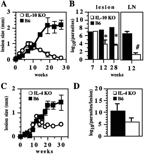
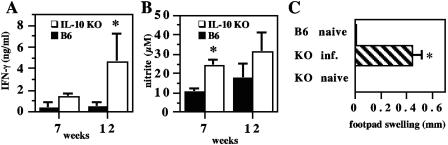
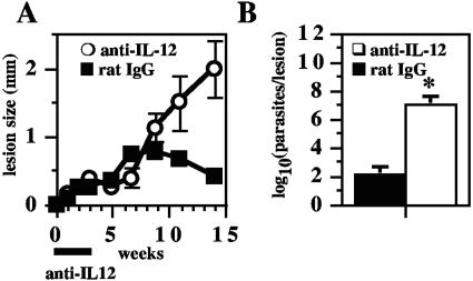
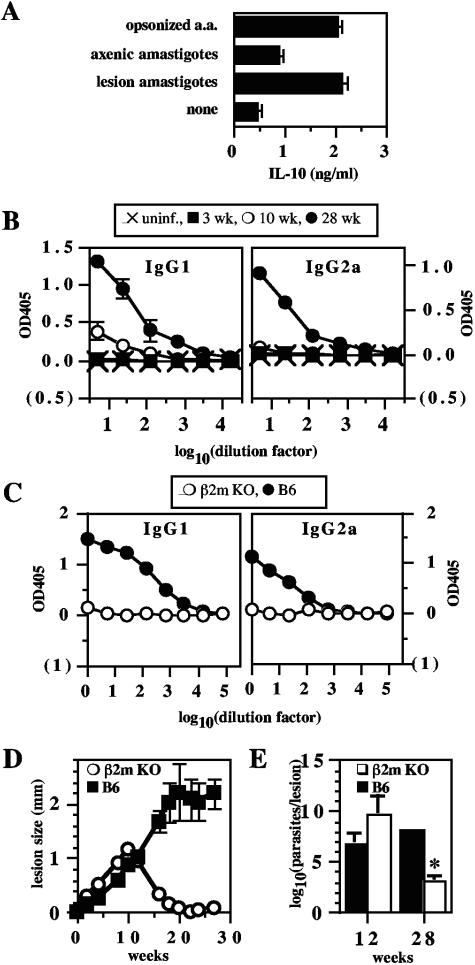
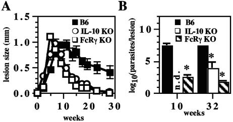
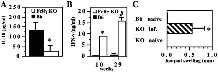
Infection of C57BL/6 (B6) mice with Leishmania mexicana is associated with a minimal immune response and chronic disease. Here we show that B6 interleukin 10-/- (IL-10-/-) mice resolve their lesions and exhibit increased gamma interferon (IFN-gamma), nitric oxide production, and delayed-type hypersensitivity. This enhanced resistance was dependent upon IL-12p40, since treatment of L. mexicana-infected IL-10-/- mice with anti-IL-12p40 monoclonal antibody abrogated healing. Antibody-opsonized L. mexicana induced IL-10 production by B6 macrophages in vitro, implicating antibody binding to Fc receptors as a mechanism involved in IL-10 production in this infection. Furthermore, B6 FcRgamma-/- mice resolve L. mexicana lesions, and lymph node cells from these mice produced less IL-10 and more IFN-gamma than cells from infected wild-type mice. These data demonstrate that removal of IL-10 or FcgammaR leads to resolution of L. mexicana disease and support a model in which ligation of FcgammaR by L. mexicana-bound immunoglobulin G promotes IL-10 production, leading to chronic disease.


Figures

FIG. 1.
FIG. 1.
IL-10−/− mice resolve L. mexicana lesions. A, IL-10−/− (IL-10 KO) and B6 mice were infected in the right hind footpad with 5 × 10⁶ stationary-phase L. mexicana promastigotes, and lesion size was monitored. B, at the times indicated, parasite burdens from IL-10−/− and B6 mice were determined by limiting dilution in the lesion and LN. *, P < 0.05; #, P < 0.001. Data are representative of two experiments with similar results. C, IL-4−/− (IL-4 KO) and B6 mice were infected and monitored as for panel A. D, at 30 weeks postinfection, lesion parasite burdens from panel C were determined by limiting dilution (P > 0.05). Data are representative of two experiments with similar results. Error bars in all figures represent SEM for groups of individual mice, except where noted.
FIG. 2.
FIG. 2.
Enhanced Th1 responses in L. mexicana-infected IL-10−/− mice. A, IL-10−/− (IL-10 KO) and B6 mice were infected with L. mexicana, and at the times indicated postinfection, draining LN cells were stimulated with FTAg for 3 days and supernatants were assayed for IFN-γ by ELISA. *, P < 0.05 for combined data of two similar experiments. No measurable IFN-γ was detectable with unstimulated media controls. B, supernatants from A were assayed for nitrite by the Griess reaction. *, P < 0.05. Data are representative of two experiments with similar results. C, DTH reactions were assessed in healed L. mexicana-infected IL-10−/− mice at 29 weeks postinfection (KO inf.) and compared with results for naive B6 and IL-10−/− mice. *, P < 0.05.
FIG. 3.
FIG. 3.
Healing of L. mexicana-infected IL-10−/− mice is mediated by IL-12p40. A, IL-10−/− mice were infected with L. mexicana as described for Fig. 1, groups were treated intraperitoneally with anti-IL-12p40 or rat IgG on days 0, 7, 14, and 21, and lesion size was monitored. B, at 18 weeks postinfection, parasite burdens from the same experiment were determined by limiting dilution. *, P = 0.003.
FIG. 4.
FIG. 4.
IgG is required for IL-10 production and chronic L. mexicana infection. A, BMMΦ from B6 mice were stimulated with 100 ng of LPS/ml alone (none) or infected with L. mexicana lesion-derived amastigotes, untreated axenic amastigotes, or opsonized axenic amastigotes (opsonized a.a.), and IL-10 levels in supernatants were measured by ELISA. P values were <0.003 for all comparisons except lesion amastigotes versus opsonized axenic amastigotes (P = 0.66). SEM are shown for quadruplicate wells. B, B6 mice were infected as described for Fig. 1. At 0 (uninf.), 3, 10, and 28 weeks postinfection, serum was collected and Leishmania-specific IgG1 and IgG2a levels were measured by ELISA. C, β2-microglubulin−/− (β2m KO) and B6 mice were infected with L. mexicana for 28 weeks as described for Fig. 1, and serum samples were assayed for Leishmania-specific IgG1 and IgG2a by ELISA. Error bars represent SEM for groups of five mice, although they are too small to see. Serum levels of Leishmania-specific IgG1 and IgG2a from β2m KO mice were indistinguishable from levels in normal mouse serum. D, β2-microglobulin−/− (β2m KO) and B6 mice were infected as described for Fig. 1, and lesion size was monitored. Data are representative of two experiments with similar results. E, At the noted times postinfection, mice were sacrificed, and footpad lesion parasite burdens were determined by limiting dilution. *, P < 0.05. Data are representative of two experiments with similar results.
FIG. 5.
FIG. 5.
FcRγ−/− mice resolve L. mexicana lesions. A, FcRγ−/− (FcRγ KO), IL-10−/− (IL-10 KO), and B6 mice were infected as described for Fig. 1, and lesion size was monitored. B, at the times indicated, lesion parasite burdens were determined by limiting dilution. *, P < 0.05 compared to B6; n.d., not done. Data are representative of two experiments with similar results.
FIG. 6.
FIG. 6.
L. mexicana-infected FcRγ−/− mice produce less IL-10 and more IFN-γ than infected control mice and develop DTH responses to rechallenge. A, B6 FcRγ−/− (FcRγ KO) and B6 mice were infected with L. mexicana for 10 weeks, draining LN cells were incubated in medium alone for 3 days, and supernatants were assayed for IL-10 by ELISA. *, P = 0.03. B, LN cells from mice infected for the indicated times with L. mexicana were restimulated for 3 days with FTAg, and supernatants were assayed for IFN-γ by ELISA. No measurable IFN-γ was detectable with unstimulated media controls. *, P < 0.05; #, P = 0.002. Data are representative of two experiments with similar results. C, DTH was assessed in healed L. mexicana-infected FcRγ−/− mice (KO inf.) at 29 weeks postinfection and compared with that in naive B6 and FcRγ−/− mice. Data are representative of two experiments with similar results. *, P < 0.05.

Similar articles

Cited by

References

    1. Afonso, L. C., and P. Scott. 1993. Immune responses associated with susceptibility of C57BL/10 mice to Leishmania amazonensis. Infect. Immun. 61:2952-2959. - PMC - PubMed
    1. Aguilar Torrentera, F., J. D. Laman, M. Van Meurs, L. Adorini, E. Muraille, and Y. Carlier. 2002. Endogenous interleukin-12 is critical for controlling the late but not the early stage of Leishmania mexicana infection in C57BL/6 mice. Infect. Immun. 70:5075-5080. - PMC - PubMed
    1. Alexander, J., F. Brombacher, H. A. McGachy, A. N. McKenzie, W. Walker, and K. C. Carter. 2002. An essential role for IL-13 in maintaining a non-healing response following Leishmania mexicana infection. Eur. J. Immunol. 32:2923-2933. - PubMed
    1. Alexander, J., G. H. Coombs, and J. C. Mottram. 1998. Leishmania mexicana cysteine proteinase-deficient mutants have attenuated virulence for mice and potentiate a Th1 response. J. Immunol. 161:6794-6801. - PubMed
    1. Alexander, J., and P. M. Kaye. 1985. Immunoregulatory pathways in murine leishmaniasis: different regulatory control during Leishmania mexicana mexicana and Leishmania major infections. Clin. Exp. Immunol. 61:674-682. - PMC - PubMed

Publication types

LinkOut - more resources