. 2005 Apr;73(4):2469-77.
doi: 10.1128/IAI.73.4.2469-2477.2005.

Q fever pneumonia: virulence of Coxiella burnetii pathovars in a murine model of aerosol infection

Affiliations

Q fever pneumonia: virulence of Coxiella burnetii pathovars in a murine model of aerosol infection

Andreas Stein et al. Infect Immun. 2005 Apr.

Abstract

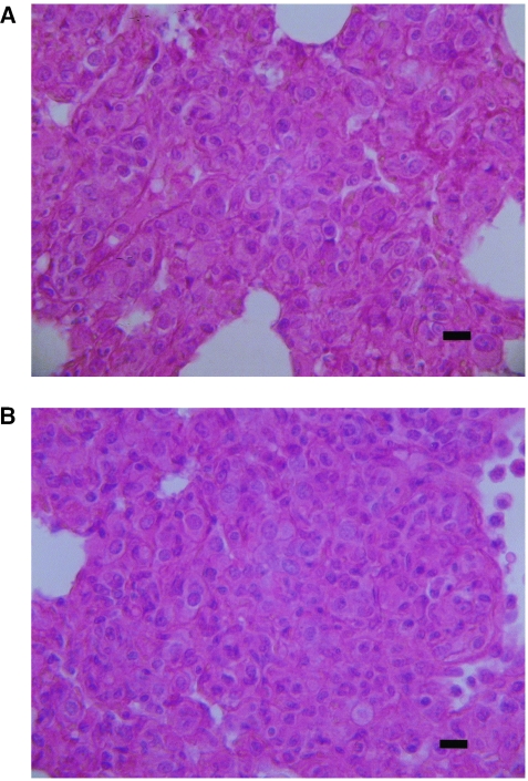
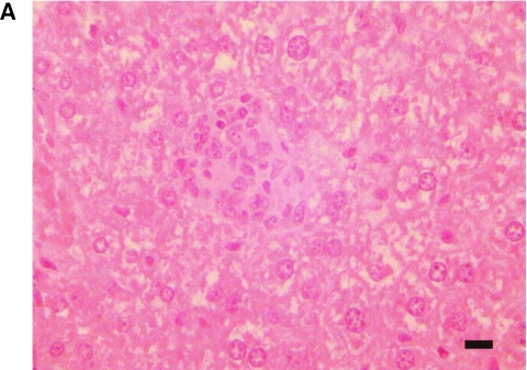
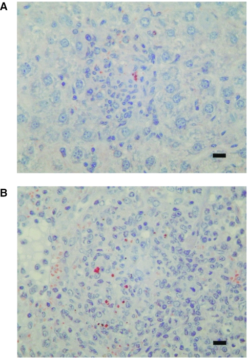
Q fever is a worldwide zoonosis caused by Coxiella burnetii, a strictly intracellular bacterium that is a potential bioweapon. Humans usually acquires Q fever after inhalation of dust infected by subclinical animals. We used an aerosol exposure apparatus to challenge immunocompetent (BALB/c) and severe combined immunodeficient (SCID) mice with two different strains (strain Nine Mile and strain Q 212) of C. burnetii at two different inocula. Pathological lesions and dissemination of the bacteria were related to the size of the inoculum. SCID mice showed major pulmonary lesions, whereas similarly infected BALB/c mice were more able to eliminate the bacteria. Pathological differences were found between the strains, with Nine Mile showing more severe histological lesions and quantified spread of bacteria. Our animal model could provide a new tool for the study of acute Q fever pneumonia, the development of Q fever in immunodeficient hosts, and the differentiation of pathogenicity among C. burnetii isolates.


Figures

FIG. 1.
FIG. 1.
C. burnetii pneumonia in mice infected with 108 Nine Mile strain organisms. The thickened alveolar walls are heavily infiltrated with mononuclear leukocytes, macrophages, and lymphocytes, with granulomatous interstitial inflammation. (A) BALB/c mouse; (B) SCID mouse. Hematoxylin-eosin-saffron staining was used. Magnification, ×400. Scale bars, 10 μm.
FIG. 2.
FIG. 2.
Sections of C. burnetii-induced granulomas at day 14 postinfection in the liver of a BALB/c mouse (A) and in the liver (B) and spleen (C) of a SCID mouse. Granulomas are located in the liver parenchyma and in the splenic red pulp. Hematoxylin-eosin was used. Magnification, ×400. Scale bars, 10 μm.
FIG. 2.
FIG. 2.
Sections of C. burnetii-induced granulomas at day 14 postinfection in the liver of a BALB/c mouse (A) and in the liver (B) and spleen (C) of a SCID mouse. Granulomas are located in the liver parenchyma and in the splenic red pulp. Hematoxylin-eosin was used. Magnification, ×400. Scale bars, 10 μm.
FIG. 3.
FIG. 3.
Demonstration of C. burnetii antigen by immunohistochemistry in the lung of a SCID mouse 7 days after infection with 108 Nine Mile strain organisms. Cytoplasmic vacuoles of macrophages in inflammatory granuloma present in interalveolar walls (A) and in alveolar air spaces (B) are packed with coarse granular immunopositive material. Polyclonal rabbit anti-C. burnetii antibody was used at a dilution of 1:2,000, along with hemalun counterstain. Magnification, ×400. Scale bars, 10 μm.
FIG. 4.
FIG. 4.
Immunohistochemical demonstration of C. burnetii antigen in liver (A) and spleen (B) of a SCID mouse at day 14 postinfection with 108 Nine Mile strain organisms. Granulomas are composed predominantly of epithelioid macrophages, in part infected. The granulomas and the splenic white pulp are almost completely devoid of lymphoid cells. Polyclonal rabbit anti-C. burnetii antibody at a dilution of 1:2,000 was used, along with hemalun counterstain. Magnification, ×400. Scale bars, 10 μm.

References

    1. Andoh, M., T. Naganawa, A. Hotta, T. Yamaguchi, H. Fukushi, T. Masegi, and K. Hirai. 2003. SCID mouse model for lethal Q fever. Infect. Immun. 71:4717-4723. - PMC - PubMed
    1. Anonymous. 1950. Experimental Q fever in man. Br. Med. J. 1:1000.
    1. Babudieri, B. 1959. Q fever: a zoonosis. Adv. Vet. Sci. Comp. Med. 5:82-182.
    1. Benson, W. W., D. W. Brock, and J. Mather. 1963. Serologic analysis of a penitentiary group using raw milk from a Q fever infected herd. Public Health Rep. 78:707-710. - PMC - PubMed
    1. Embil, J., J. C. Williams, and T. J. Marrie. 1990. The immune response in a cat-related outbreak of Q fever as measured by the indirect immunofluorescence test and the enzyme-linked immunosorbent assay. Can. J. Microbiol. 36:292-296. - PubMed

LinkOut - more resources