. 2005 Apr;73(4):2524-32.
doi: 10.1128/IAI.73.4.2524-2532.2005.

Shiga toxin is transported from the endoplasmic reticulum following interaction with the luminal chaperone HEDJ/ERdj3

Affiliations

Shiga toxin is transported from the endoplasmic reticulum following interaction with the luminal chaperone HEDJ/ERdj3

Min Yu et al. Infect Immun. 2005 Apr.

Abstract

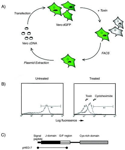
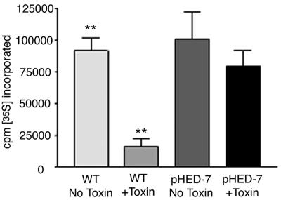
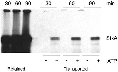
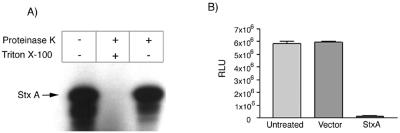
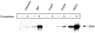
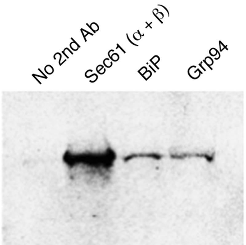
Shiga toxin (Stx) follows a complex intracellular pathway in order to kill susceptible cells. After binding to cell surface glycolipids, the toxin is internalized and trafficked in retrograde fashion to the endoplasmic reticulum (ER). From the ER lumen, the toxin must gain access to the cytoplasm, where it enzymatically inactivates the 28S rRNA, inhibiting protein synthesis. The host molecules involved in this pathway and the mechanisms utilized by the toxin to access the cytoplasm from the ER are largely unknown. We found that Stx is capable of energy-dependent transport across the ER lumen, as has recently been demonstrated for the cholera and ricin toxins. Genetic screening for molecules involved in Shiga toxin trafficking yielded a cDNA encoding a prematurely truncated protein. Characterization of this cDNA revealed that it encodes a novel Hsp40 chaperone, designated HEDJ or ERdj3, localized to the ER lumen, where it interacts with BiP, a molecule known to be involved in protein retrotranslocation out of the ER. We demonstrated that within the ER lumen Stx interacts with HEDJ and other chaperones known to be involved in retrotranslocation of proteins across the ER membrane. Moreover, sequential immunoprecipitation revealed that Shiga toxin was present in a complex that included HEDJ and Sec61, the translocon through which proteins are retrotranslocated to the cytoplasm. These findings suggest that HEDJ is a component of the ER quality control system and that Stx utilizes HEDJ and other ER-localized chaperones for transport from the ER lumen to the cytosol.


Figures

FIG. 1.
FIG. 1.
(A) Schematic drawing of the phenotypic cloning approached used to isolate HEDJ. Vero cells stably expressing destabilized green fluorescent protein (Vero:dGFP) were transfected with a Vero cell cDNA library and then exposed to toxin. After 18 h of incubation, toxin-resistant cells were separated from susceptible cells by FACS. Plasmid DNA was recovered from enriched cells by Hirt extraction and were subjected to another two rounds of transfection and enrichment. (B) FACS profile demonstrating the baseline fluorescence of untreated Vero:dGFP cells and cells treated for 18 h either with cycloheximide or with Shiga toxin. The region gated by M1 included less than 1% Vero cells lacking green fluorescent protein expression. (C) Schematic representation of the HEDJ gene product. The residues enriched by phenotypic cloning and included in plasmid pHED-7 are indicated by the dotted line. The locations of the J domain, the glycine- and phenylalanine-rich domain (G/F region), and the cysteine-rich domain found in most Hsp40 chaperones (36) are indicated.
FIG. 2.
FIG. 2.
Cells transfected with pHED-7 exhibit reduced susceptibility to Shiga toxin. Vero cells were transfected with either plasmid alone (WT) or plasmid a containing truncated HEDJ cDNA (pHED-7). Neomycin-resistant cells were selected for 2 weeks and then exposed to Shiga toxin for 4 h. The cells were pulsed with [35S]methionine for 30 min in methionine-free medium. After several washes, the cells were lysed, TCA was added to a concentration of 10%, and the amount of radioactivity incorporated into protein was quantified by scintillation counting of the TCA precipitate. Two asterisks indicate that the difference in [35S]methionine incorporation between untreated and toxin-treated wild-type cells is statistically significant (P < 0.05).
FIG. 3.
FIG. 3.
StxA is transported across the ER membrane in an ATP-dependent manner. StxA was synthesized by coupled in vitro transcription and translation in the presence of ER extracts and 35S-labeled methionine and cysteine as described previously. Microsomes were then washed in PBS and resuspended in buffer lacking ATP or containing ATP and an ATP-regenerating system. After 30, 60, or 90 min of incubation at 37°C, microsomes were separated from the supernatant, and all samples were boiled and subjected to SDS-PAGE. The toxin remaining in the microsomes (Retained) and the toxin transported into the supernatant (Transported) were detected by fluorography.
FIG. 4.
FIG. 4.
Demonstration that StxA is incorporated into the ER lumen and is active. (A) StxA was synthesized by coupled in vitro transcription and translation in the presence of 35S-labeled methionine and cysteine and ER extracts. The microsomal extract was aliquoted into three tubes and exposed either to no proteinase K or detergent, to both proteinase K and detergent, or to proteinase K without detergent. After 30 min of incubation, the reaction mixtures were subjected to SDS-PAGE and fluoroscopy to detect residual toxin. (B) StxA was synthesized by coupled in vitro transcription and translation in the presence of ER extracts. Microsomes were then permeabilized with 0.1% Triton X-100 and diluted 100-fold with PBS, and 1-μl portions were added to rabbit reticulocyte lysates. As a control, an in vitro transcription-translation reaction mixture containing empty expression plasmid was used as the microsome source. After reticulocyte lysates were treated with Stx or control extracts, a plasmid encoding luciferase was added, and a second in vitro transcription and translation reaction was carried out. The amount of luciferase produced was quantified by adding 5 μl of the translation reaction mixture to luciferase substrate, and the light output was quantified with a luminometer. RLU, relative light units.
FIG. 5.
FIG. 5.
StxA coimmunoprecipitates with HEDJ and other luminal ER chaperones. StxA was synthesized by coupled in vitro transcription and translation in the presence of 35S-labeled methionine and cysteine and ER extracts. Microsomes were aliquoted and either exposed to a cross-linker (DSP) (+) or not cross-linked (−). The microsomes were then washed and subjected to immunoprecipitation with antibodies against various ER chaperones. The immunoprecipitated material was boiled and subjected to SDS-PAGE. Coimmunoprecipitated toxin was detected by fluorography.
FIG. 6.
FIG. 6.
StxA is found in a complex that includes HEDJ and Sec61. StxA was synthesized by coupled in vitro transcription and translation in the presence of 35S-labeled methionine and cysteine and ER extracts as described previously. After cross-linking with DSP, the first immunoprecipitation was carried out with antibodies against HEDJ. The immunoprecipitated material was heated to 95°C for 5 min and then subjected to a second round of immunoprecipitation with either no second antibody (Ab) or antibody against Sec61, BiP, or Grp94.

References

    1. Bies, C., S. Guth, K. Janoschek, W. Nastainczyk, J. Volkmer, and R. Zimmermann. 1999. A Scj1p homolog and folding catalysts present in dog pancreas microsomes. Biol. Chem. 380:1175-1182. - PubMed
    1. Elliott, S. P., M. Yu, H. Xu, and D. B. Haslam. 2003. Forssman synthetase expression results in diminished Shiga toxin susceptibility: a role for glyocolipids in determining host-microbe interactions. Infect. Immun. 71:6543-6552. - PMC - PubMed
    1. Garred, O., B. van Deurs, and K. Sandvig. 1995. Furin-induced cleavage and activation of Shiga toxin. J. Biol. Chem. 270:10817-10821. - PubMed
    1. Hazes, B., and R. J. Read. 1997. Accumulating evidence suggests that several AB-toxins subvert the endoplasmic reticulum-associated protein degradation pathway to enter target cells. Biochemistry 36:11051-11054. - PubMed
    1. Hirt, B. 1967. Selective extraction of polyoma DNA from infected mouse cell cultures. J. Mol. Biol. 26:365-369. - PubMed

Publication types

LinkOut - more resources