. 2005 Apr;73(4):2533-40.
doi: 10.1128/IAI.73.4.2533-2540.2005.

Designer arrays for defined mutant analysis to detect genes essential for survival of Mycobacterium tuberculosis in mouse lungs

Affiliations

Designer arrays for defined mutant analysis to detect genes essential for survival of Mycobacterium tuberculosis in mouse lungs

Gyanu Lamichhane et al. Infect Immun. 2005 Apr.

Abstract

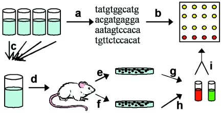
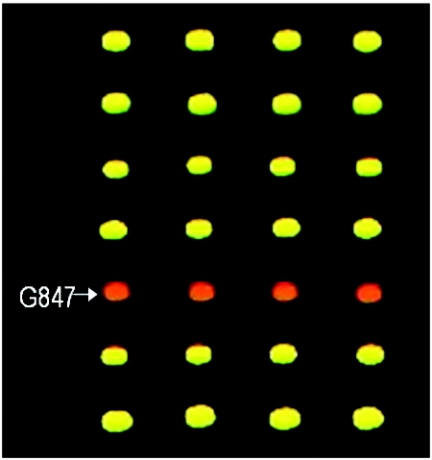
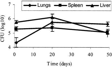
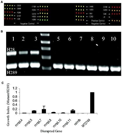
The mechanisms by which Mycobacterium tuberculosis elicits disease are complex, involving a large repertoire of bacterial genes that are required for in vivo growth and survival. To identify such genes, we utilized a high-throughput microarray detection method to rapidly screen hundreds of unique, genotypically defined transposon mutants for in vivo survival with a high degree of specificity and sensitivity. Thirty-one M. tuberculosis genes were found to be required for in vivo survival in mouse lungs. These genes are involved in a broad range of activities, including metabolism, cell wall functions, and regulation. Our screen included 11 of the 12 known members of the mycobacterial membrane protein (mmpL) family genes, and mutation of 6 of these genes-mmpL4, mmpL5, mmpL7, mmpL8, mmpL10, and mmpL11-severely compromised the ability of the mutants to multiply in mouse lungs. Most of the 31 genes are conserved in other pathogenic mycobacteria, including M. leprae and M. bovis, suggesting that a core of basic in vivo survival mechanisms may be highly conserved despite the divergent human pathology caused by members of the mycobacterial genus. Of the 31 genes reported here, 17 have not been previously described to be involved in in vivo growth and survival of M. tuberculosis.


Figures

FIG. 1.
FIG. 1.
Schematics of DeADMAn. (Step a) Mutants are grown individually in liquid culture, and the sequence of the transposon insertion junction is determined. (Step b) 60-mer oligonucleotides corresponding to insertion junction sequence of each mutant are printed on a custom microarray. (Step c) The mutants are pooled. (Step d) Mice are infected with pooled mutants to determine in vivo growth. (Steps e and f) The input pool is obtained by harvesting lungs and spleen 1 day postinfection (e) and the output pool is obtained by harvesting lungs and spleen at 20 and 49 days postinfection (f). (Steps g and h) Probes are prepared from the input pool sample (g) and probes are prepared from the output pool sample (h). (Step i) The input and output probes, which are labeled with Cy5 (red) and Cy3 (green) dyes, are cohybridized to custom microarray, and the relative levels of probes corresponding to each mutant are determined. A mutant attenuated for in vivo growth would be absent or underrepresented in the output pool, and this phenotype would be revealed by overabundance of the Cy5-labeled input probe producing red spots. Spots that appear yellow-orange result from the presence and cohybridization of both the input (Cy5) and output (Cy3) probes.
FIG. 2.
FIG. 2.
Growth-attenuated mutants are readily detected by DeADMAn. A subsection of the array for pool C is shown. Probe from the input pool at day 1 postinfection was labeled with Cy5 (red), and the probe from the output pool at day 49 postinfection was labeled with Cy3 (green). The mutant, G847, which has a mutation in the gene bioB, is underrepresented in the output pool.
FIG. 3.
FIG. 3.
Growth in vivo of pools of mutants is similar to standard growth of wild-type M. tuberculosis. Mice were infected with a pool of 100 different mutants, i.e., pool A. Lungs, spleens, and liver were obtained at days 1, 20, and 49 postinfection, and the number of viable bacilli quantified by plating on solid medium.
FIG. 4.
FIG. 4.
(A) Microarray with input probes labeled with Cy5 (red) and output probes labeled with Cy3 (green) that were prepared by using colonies obtained from the lungs of mice. Each oligonucleotide represents a junction sequence for a mutant and is printed four times. The mutants that were underrepresented in the output pool (attenuated for in vivo survival) have proportionately more Cy5 probe and produce red spots on the array. There are four controls on this array: (i) H28 is a mutant from a different pool, pool A, that was observed to be attenuated and was used in this pool as a control; (ii) H289 is another mutant from pool A that was not attenuated; (iii) spots labeled as positive controls contain a mixture of all of the oligonucleotides printed on the arrays and thus is an internal positive control; and (iv) spots labeled as negative controls are oligonucleotides representing four different mutants from a different pool and thus serve as internal controls for background noise. (B) Agarose gel electrophoresis showing PCR amplicons of junctions of H28 and H289 (control). Lanes 1, 2, and 3 represent mutants recovered from lungs of three mice sacrificed at 1 day postinfection (input pool). Similarly, lanes 5, 6 and 7, and 8, 9, and 10 represent mutants recovered from three mice sacrificed at 49 and 98 days postinfection, respectively. Lane 4 is 1-kb plus marker (Invitrogen). (C) Six mmpL mutants and a control, H28, are compared against H289 for growth in vivo. Mutants H28 and H289 were used as controls based on their phenotypes from an earlier experiment. H28 is one of the 100 mutants of pool A and was observed to be attenuated. H289 is another mutant from pool A that was not attenuated for growth in vivo. Growth index refers to the population of each mutant, as measured from signals from the DeADMAn arrays, in the lungs of mice relative to the population of the positive control strain H289. The errors bars represent the standard deviation.

References

    1. Akerley, B. J., E. J. Rubin, A. Camilli, D. J. Lampe, H. M. Robertson, and J. J. Mekalanos. 1998. Systematic identification of essential genes by in vitro mariner mutagenesis. Proc. Natl. Acad. Sci. USA 95:8927-8932. - PMC - PubMed
    1. Ausubel, F. M., R. Brent, R. E. Kingston, D. D. Moore, J. G. Seidman, J. A. Smith, and K. Struhl. 1998. Current protocols and molecular biology. John Wiley & Sons, New York, N.Y.
    1. Avarbock, D., J. Salem, L. S. Li, Z. M. Wang, and H. Rubin. 1999. Cloning and characterization of a bifunctional RelA/SpoT homologue from Mycobacterium tuberculosis. Gene 233:261-269. - PubMed
    1. Camacho, L. R., P. Constant, C. Raynaud, M. A. Laneelle, J. A. Triccas, B. Gicquel, M. Daffe, and C. Guilhot. 2001. Analysis of the phthiocerol dimycocerosate locus of Mycobacterium tuberculosis: evidence that this lipid is involved in the cell wall permeability barrier. J. Biol. Chem. 276:19845-19854. - PubMed
    1. Camacho, L. R., D. Ensergueix, E. Perez, B. Gicquel, and C. Guilhot. 1999. Identification of a virulence gene cluster of Mycobacterium tuberculosis by signature-tagged transposon mutagenesis. Mol. Microbiol. 34:257-267. - PubMed

Publication types

MeSH terms

Substances

LinkOut - more resources