

Detection of entorhinal layer II using 7Tesla [corrected] magnetic resonance imaging
- PMID: 15786476
- PMCID: PMC3857582
- DOI: 10.1002/ana.20426
Detection of entorhinal layer II using 7Tesla [corrected] magnetic resonance imaging
Erratum in
- Ann Neurol. 2005 Jul;58(1):172
Abstract
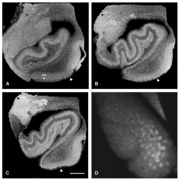
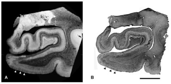
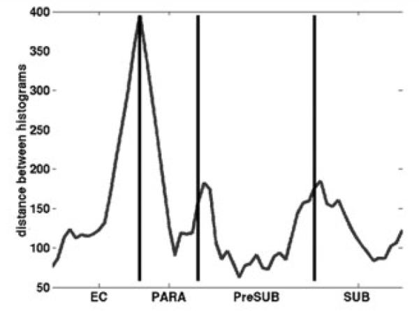
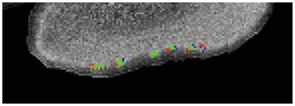
The entorhinal cortex lies in the mediotemporal lobe and has major functional, structural, and clinical significance. The entorhinal cortex has a unique cytoarchitecture with large stellate neurons in layer II that form clusters. The entorhinal cortex receives vast sensory association input, and its major output arises from the layer II and III neurons that form the perforant pathway. Clinically, the neurons in layer II are affected with neurofibrillary tangles, one of the two pathological hallmarks of Alzheimer's disease. We describe detection of the entorhinal layer II islands using magnetic resonance imaging. We scanned human autopsied temporal lobe blocks in a 7T human scanner using a solenoid coil. In 70 and 100 microm isotropic data, the entorhinal islands were clearly visible throughout the anterior-posterior extent of entorhinal cortex. Layer II islands were prominent in both the magnetic resonance imaging and corresponding histological sections, showing similar size and shape in two types of data. Area borders and island location based on cytoarchitectural features in the mediotemporal lobe were robustly detected using the magnetic resonance images. Our ex vivo results could break ground for high-resolution in vivo scanning that could ultimately benefit early diagnosis and treatment of neurodegenerative disease.
Figures
References
-
- Cajal RS. Studies in the cerebral cortex. In: Kraft LM, translator. Yearbook. Chicago: 1955. pp. 164–179.
-
- Lorente de No R. Studies on the structure of the cerebral cortex. Journal Fur Psychologie and Neurologie. 1933;45:381–438.
-
- Insausti R, Tunon T, Sobreviela T, et al. The human entorhinal cortex: a cytoarchitectonic analysis. J Comp Neurol. 1995;355:171–198. - PubMed
-
- Solodkin A, Van Hoesen GW. Entorhinal cortex modules of the human brain. J Comp Neurol. 1996;365:610–617. - PubMed
-
- Retzius G. Das Menschenhirn. Norstedt and Sonhe; Stockholm: 1896.
Publication types
MeSH terms
Grants and funding
- R01 NS070963/NS/NINDS NIH HHS/United States
- P41-RR14075/RR/NCRR NIH HHS/United States
- S10 RR019307/RR/NCRR NIH HHS/United States
- U54 EB005149/EB/NIBIB NIH HHS/United States
- P41 RR014075/RR/NCRR NIH HHS/United States
- R01 NS052585/NS/NINDS NIH HHS/United States
- T32 EB001680/EB/NIBIB NIH HHS/United States
- R01 RR16594-01A1/RR/NCRR NIH HHS/United States
- K01 AG028521/AG/NIA NIH HHS/United States
- R01 RR016594/RR/NCRR NIH HHS/United States
- R21 NS072652/NS/NINDS NIH HHS/United States
- P41 RR006009/RR/NCRR NIH HHS/United States
- R01 EB006758/EB/NIBIB NIH HHS/United States
- P41 EB015896/EB/NIBIB NIH HHS/United States
- S10 RR023401/RR/NCRR NIH HHS/United States
LinkOut - more resources
Full Text Sources
Medical
