. 2005 Jun;16(6):3064-76.
doi: 10.1091/mbc.e05-02-0174. Epub 2005 Mar 23.

Roles of polymerization dynamics, opposed motors, and a tensile element in governing the length of Xenopus extract meiotic spindles

Affiliations

Roles of polymerization dynamics, opposed motors, and a tensile element in governing the length of Xenopus extract meiotic spindles

T J Mitchison et al. Mol Biol Cell. 2005 Jun.

Abstract

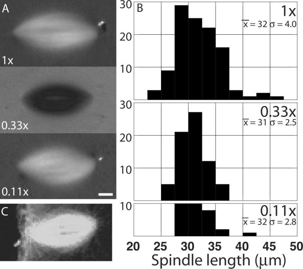
Metaphase spindles assemble to a steady state in length by mechanisms that involve microtubule dynamics and motor proteins, but they are incompletely understood. We found that Xenopus extract spindles recapitulate the length of egg meiosis II spindles, by using mechanisms intrinsic to the spindle. To probe these mechanisms, we perturbed microtubule polymerization dynamics and opposed motor proteins and measured effects on spindle morphology and dynamics. Microtubules were stabilized by hexylene glycol and inhibition of the catastrophe factor mitotic centromere-associated kinesin (MCAK) (a kinesin 13, previously called XKCM) and destabilized by depolymerizing drugs. The opposed motors Eg5 and dynein were inhibited separately and together. Our results are consistent with important roles for polymerization dynamics in regulating spindle length, and for opposed motors in regulating the relative stability of bipolar versus monopolar organization. The response to microtubule destabilization suggests that an unidentified tensile element acts in parallel with these conventional factors, generating spindle shortening force.


Figures

Figure 1.
Figure 1.
Length of in vitro spindles is independent of spindle density. Cycled spindles were assembled at a final density of 50, 16.7, and 5.5 sperm nuclei/μl and imaged in oil overlay chambers by polarization microscopy 90–120 min after addition of cytostatic factor (CSF) extract. (A) Representative images at each dilution. Bar, 5 μm. (B) Histograms of spindle lengths at each dilution measured from polarization images. The mean and SD (in micrometers) is written on each histogram. Note they are similar at each dilution. (C) Immunofluorescence image of the meiosis II spindle in an unfertilized egg, at a comparable magnification, for comparison. The animal cortex of the egg is to the left. Anti-tubulin staining and laser confocal imaging. Image kindly provided by David Gard (University of Utah) (see Cha et al., 1998).
Figure 2.
Figure 2.
Hexylene glycol promotes spindle expansion. Hexylene glycol [2% (vol/vol)] was added to cycled spindles from a 20% stock in water immediately before imaging. (A) Polarization images from a time-lapse sequence. Elapsed time shown in minutes and seconds. Hexylene glycol was added ∼1 min before the first time point. Note the growth in spindle length and width. Time elapsed shown as minutes:seconds. Length of time box, 10 μm. See Movie M1 (B) Spindle length as a function of time after addition of hexylene glycol alone (2%; black circles), hexylene glycol plus AMPPNP (1.5 mM; open squares), or hexylene glycol plus monastrol (400 μM) and p50 dynamitin (0.9 mg/ml; gray diamonds). (C) Spinning disk confocal fluorescence images of a spindle containing speckle level tubulin to which 2% hexylene glycol has been added. The line in the second panel was used to make the kymograph in D. See Movie M2. Bar, 5 μm. (D) Kymograph through the upper pole of the spindle shown in C. Time is vertical, distance along the line horizontal. Note the movement of the pole away from the spindle equator, which is to the right in the panel. Note also that many thin lines, which are speckle trajectories, parallel the movement of the pole (black lines highlight examples). Near the equator, speckles also are moving in the other direction (white line), in parallel with the opposite pole.
Figure 3.
Figure 3.
Titration of anti-MCAK antibody effects. Affinity-purified, inhibitory antibody to MCAK (Walczak et al., 1996) was added to spindle assembly reactions containing X-rhodamine tubulin 20 min after bring the extract back into M phase after replication, well before spindle assembly. Samples were squash-fixed at 80 and 120 min after bringing the extract into M phase. (A) Representative spindles fixed at 80 min as a function of final antibody concentration. Note the massive microtubule growth out of the spindle at 15 μg/ml (and higher; our unpublished data), indicative of transition to unbounded dynamic instability. Bar, 10 μm. (B) Quantification of spindle length in this experiment. Open bars are 80- and black 120-min samples. The error bars indicate one SD above and below the mean, and the number in each bar the sample size. (C) Quantification of total microtubules per spindle measured as integrated fluorescence intensity minus local background signal. Labeling as in B.
Figure 4.
Figure 4.
Inhibition of MCAK promotes microtubule outgrow and pole curling, but not spindle elongation. Affinity-purified, inhibitory antibody to MCAK was added at 150 μg/ml final to cycled spindles immediately before imaging. (A) Polarization images from a time-lapse sequence. Note the dramatic outgrowth of microtubules from the spindle (arrows in second panel). There is some alteration of spindle shape and pole structure over the sequence, but pole-to-pole distance remains approximately constant. Elapsed times shown in minutes:seconds. Antibody was added ∼1 min before the first time point. Time bar, 18 μm. See Movie M3. (B) Spinning disk confocal fluorescence images of a spindle containing speckle level X-rhodamine tubulin (green) and Alexa488-labeled antibodies to NUMA at 5 μg/ml as a pole marker (red). Note that the upper pole, visualized by anti-NUMA localization (white arrow), is curled over and apparently attached to the body of the spindle. By the last time point, the pole has moved toward the spindle equator, and become partially disorganized. In the tubulin channel, the movements of the microtubules that are associated with this pole movement can be visualized (blue arrows). In the center of the spindle, microtubules slide apart in both directions. In the upper part, most of the flow is upwards. At the top of the spindle, the flow curls over, correlating with movement of the pole back toward the equator. Bar, 5 μm. See Movie M4.
Figure 5.
Figure 5.
Rapid microtubule depolymerization by using nocodazole. Cycled spindles containing labeled tubulin (green) and a kinetochore marker (anti-CenpA; red) were mixed with nocodazole (20 μM final) and wide-field time-lapse imaging was initiated ∼10 s after mixing by using a 40× dry objective. (A) Imaging of spindle collapse. Elapsed time is shown in seconds relative to the first image; tubulin fluorescence is normalized to peak brightness in each image, to highlight the organization of remaining microtubules. Insets show 3× magnification of marked kinetochores. Note the sister kinetochores are initially well separated. The spindle rapidly looses fluorescence, shortens, and the sister kinetochore move together. Note some evidence of buckling of kinetochore microtubules at 59, 119 s. Bar, 5 μm (main panels), 1.7 μm (insets). See Movie M5. (B) Quantification of the sequence in A, showing distance between the marked kinetochores in A (k-k), pole to pole distance (p-p) and integrated tubulin fluorescence after subtracting local background signal. Note that k-k distance drops faster (in percent terms) than p-p distance.
Figure 6.
Figure 6.
Microtubule depolymerization using a caged drug. Cycled spindles containing labeled tubulin, kinetochore, and pole markers in some cases, and caged 105D were imaged by time-lapse wide-field (B and C) or confocal (D) fluorescence. At t = 0, 105D was photoreleased by 1- to 2-s illumination with a UV filter set. All times are in seconds relative to photorelease. Bar, 7.5 μm (B), 6 μm (C), 5 μm (D), and 1.25 μm (D, insets). (A) Structure of caged 105D and the photochemical reaction that releases active drug. (B) Example of a spindle before and after photorelease of 105D (wide-field). Tubulin fluorescence is presented with brightness and contrast held constant throughout the sequence to highlight the rapid decrease in microtubule density. This sequence also contained a fluorescent kinetochore probe; see Movie M6. (C) Second widefield example. Tubulin fluorescence is normalized to peak brightness in each image, to highlight the organization of remaining microtubules. Note the decrease in spindle length, and buckling of remaining stable microtubules. See Movie M7. (D) Example of photorelease of 105D (confocal). Probes are for tubulin (green), kinetochore (anti-CenpA; red), and poles (anti-NUMA; red). Tubulin fluorescence is normalized to peak brightness in each image. Insets show marked kinetochore pairs at 4× magnification. The focal plane was changed several times during this sequence, and different kinetochore pairs are shown in each panel. At late time points, most of the remaining stable microtubules connect to kinetochores. Note that the poles (large red dots) move progressively together. Sister kinetochores first move together and then twist and move away from the spindle axis, whereas their attached microtubules either buckle or move outward, suggesting kinetochore fibers experience compression from the collapsing poles. The spindle was optically sectioned twice during this sequence, and the images at 941 and 978 s are two focal planes from one through-focal series. Note the absence of straight microtubule bundles directly connecting the poles at any time point or focal plane.
Figure 7.
Figure 7.
Eg5 and dynein/dynactin play antagonistic roles in spindle assembly. Cycled spindles were assembled in the presence of no drug, the dynactin inhibitor p50 dynamitin (0.9 mg/ml), the Eg5 inhibitor monastrol (200 μM), or both. Drugs were added at the time of CSF addition, and spindles were imaged live in oil overlay chambers by polarization microscopy 90–120 min later. A panel of representative images is shown for each condition. Note the expected appearance of p50 spindles with unfocussed poles and monopolar monastrol spindles. When both drugs were added, bipolarity was completely rescued, and pole focusing was partially rescued. Bar, 10 μm. See Table 1 for quantitation.
Figure 8.
Figure 8.
Antagonizing dynein/dynactin rescues the effects of Eg5 depeletion. Extracts were depleted of Eg5 by using affinity-purified antibody and magnetic beads. Three rounds of depletion were used to remove all Eg5 that could be detected on Western blots (>95%). (A) Western blot analysis of Eg5 and mock-depleted extracts. Eg5 is removed to below the detection limit in the depleted extract. (B) Quantitation of spindle morphology. Eg5 and mock-depleted extracts were used to assemble cycled spindles in the presence or absence of p50 dynamitin (0.9 mg/ml). In Eg5 depletion alone, the majority of spindles were monopolar asters. Addition of p50 promoted assembly of mostly bipolar spindles. (C) Typical spindle assembled in mock depleted extract. (D) Typical spindle assembled in mock-depleted extract + p50. Note bipolar organization with splayed poles. (E) Typical spindle-assembled in Eg5-depleted extract. Note monopolar organization. (F) Typical spindle assembled in Eg5 depleted extract + p50. Note rescue of both bipolarity and pole morphology. Bar, 5 μm.
Figure 9.
Figure 9.
Interpretation of results. See text for details.

References

    1. Cassimeris, L., Rieder, C. L., Rupp, G., and Salmon, E. D. (1990). Stability of microtubule attachment to metaphase kinetochores in PtK1 cells. J. Cell Sci. 96, 9-15. - PubMed
    1. Cha, B. J., Error, B., and Gard, D. L. (1998). XMAP230 is required for the assembly and organization of acetylated microtubules and spindles in Xenopus oocytes and eggs. J. Cell Sci. 111, 2315-2327. - PubMed
    1. Chang, P., Jacobsen, M. K., and Mitchison, T. J. (2004). Poly(ADP-ribose) is required for spindle assembly and structure. Nature 432, 645-649. - PubMed
    1. Cytrynbaum, E. N., Scholey, J. M., and Mogilner, A. (2003). A force balance model of early spindle pole separation in Drosophila embryos. Biophys. J. 84, 757-769. - PMC - PubMed
    1. Desai, A., Murray, A., Mitchison, T. J., and Walczak, C. E. (1999a). The use of Xenopus egg extracts to study mitotic spindle assembly and function in vitro. Methods Cell Biol. 61, 385-412. - PubMed

Publication types