. 2005 Jun;16(6):2836-47.
doi: 10.1091/mbc.e04-10-0926. Epub 2005 Mar 23.

The Xenopus TACC homologue, maskin, functions in mitotic spindle assembly

Affiliations

The Xenopus TACC homologue, maskin, functions in mitotic spindle assembly

Lori L O'Brien et al. Mol Biol Cell. 2005 Jun.

Abstract

Maskin is the Xenopus homolog of the transforming acidic coiled coil (TACC)-family of microtubule and centrosome-interacting proteins. Members of this family share a approximately 200 amino acid coiled coil motif at their C-termini, but have only limited homology outside of this domain. In all species examined thus far, perturbations of TACC proteins lead to disruptions of cell cycle progression and/or embryonic lethality. In Drosophila, Caenorhabditis elegans, and humans, these disruptions have been attributed to mitotic spindle assembly defects, and the TACC proteins in these organisms are thought to function as structural components of the spindle. In contrast, cell division failure in early Xenopus embryo blastomeres has been attributed to a role of maskin in regulating the translation of, among others, cyclin B1 mRNA. In this study, we show that maskin, like other TACC proteins, plays a direct role in mitotic spindle assembly in Xenopus egg extracts and that this role is independent of cyclin B. Maskin immunodepletion and add-back experiments demonstrate that maskin, or a maskin-associated activity, is required for two distinct steps during spindle assembly in Xenopus egg extracts that can be distinguished by their response to "rescue" experiments. Defects in the "early" step, manifested by greatly reduced aster size during early time points in maskin-depleted extracts, can be rescued by readdition of purified full-length maskin. Moreover, defects in this step can also be rescued by addition of only the TACC-domain of maskin. In contrast, defects in the "late" step during spindle assembly, manifested by abnormal spindles at later time points, cannot be rescued by readdition of maskin. We show that maskin interacts with a number of proteins in egg extracts, including XMAP215, a known modulator of microtubule dynamics, and CPEB, a protein that is involved in translational regulation of important cell cycle regulators. Maskin depletion from egg extracts results in compromised microtubule asters and spindles and the mislocalization of XMAP215, but CPEB localization is unaffected. Together, these data suggest that in addition to its previously reported role as a translational regulator, maskin is also important for mitotic spindle assembly.


Figures

Figure 1.
Figure 1.
Maskin localizes to microtubule structures in egg extracts. (A) Western blot to characterize our maskin antibody. rec, recombinant maskin; ext, extract. Molecular-weight markers are indicated on the left. (B) Western blot to titrate the amount of maskin present in egg extract. The amount of egg extract and recombinant maskin of known concentration loaded in each lane is indicated. The concentration of maskin depends on the egg extract and ranges between 10 and 20 nM (4 independent measurements using 4 different extracts); maskin concentration is ∼14 nM in the extract shown (total protein concentration in this extract is ∼50 mg/ml). (C) Maskin (green) localizes along microtubules (red) in asters and spindles induced by addition to the egg extract of GST-RanL43E (top row) or sperm chromatin (bottom rows; DNA is blue in the overlays). Scale bar, 20 μm.
Figure 2.
Figure 2.
Maskin depletion disrupts microtubule aster and spindle assembly in Xenopus egg extracts. (A) Fluorescence micrographs of microtubule asters (formed within 15 min of incubation) assembled in mock-depleted, maskin-depleted, or maskin-depleted plus bacterially expressed purified recombinant protein (as indicated). Amino acids 714–931 of maskin constitute the TACC domain of maskin (see Figure 7 for schematic). Recombinant proteins were expressed as GST fusion proteins, but GST was cleaved off following purification. Top row, tubulin staining; microtubules are red and DNA is blue in the overlays (bottom row). Scale bar, 25 μm. (B) Quantitation of microtubule fluorescence intensity of asters assembled in depleted extracts. The graph represents the average of three independent experiments; the error bars represent the SD. (C) Examples of normal and abnormal spindles (formed after 90 min of incubation) in maskin-depleted extracts, as indicated. Microtubules are red and DNA is blue in the overlays. Scale bar, 25 μm. (D) Quantitation of the ratio of normal and abnormal spindles in mock- and maskin-depleted extracts, and in maskin-depleted extracts reconstituted with recombinant full-length maskin, as indicated. The graph represents the average of three independent experiments.
Figure 3.
Figure 3.
Maskin depletion has little effect on γ-tubulin recruitment to centrosomes. (A) Fluorescence micrographs of sperm chromatin incubated in mock-depleted (top rows) or maskin-depleted (bottom rows) egg extract supplemented with nocodazole to prevent microtubule formation. Acetylated tubulin (a marker for centrioles) is red, γ-tubulin is green, and DNA is blue in the overlays (right panels). Scale bar, 5 μm. (B) Quantitation of the amount of γ-tubulin recruited to sperm centrosomes. To quantitate the amount of γ-tubulin recruited, the γ-tubulin fluorescence intensity associated with the sperm centrioles was measured using MetaMorph software. The graph represents the average of 11 independent experiments ± SEM.
Figure 4.
Figure 4.
Maskin is involved in centrosome-independent microtubule assembly in Xenopus egg extracts. (A) Ran-aster formation in mock- and maskin-depleted extracts. Fluorescence micrographs of microtubule structures assembled in mock-depleted (top left-hand panel), maskin-depleted (top right-hand panel), maskin-depleted plus bacterially expressed recombinant maskin (bottom left-hand panel), or mock-depleted plus an extra bolus of bacterially purified recombinant maskin (bottom right-hand panel). Scale bar, 20 μm. (B) Quantitation of Ran-induced structures assembled in depleted extracts. Graph on the left, average fluorescence intensity; graph on the right, average microtubule length; mock, mock-depleted; depl, maskin-depleted; recon, maskin-depleted extract plus 20 nM bacterially purified recombinant maskin; extra, mock-depleted extract plus 20 nM bacterially purified recombinant maskin. (C) Western blot to quantitate the levels of maskin depletion and add-back, as indicated.
Figure 5.
Figure 5.
Maskin is a microtubule-binding protein. (A) Coomassie-stained gel of the supernatant (S) and pellets (P) of a representative microtubule copelleting assay using a constant amount of maskin and increasing amounts of taxol-stabilized microtubules, as indicated below the gel. (B) Plot of the percentage of maskin associated with microtubules versus the tubulin concentration. The data shown are the averages (±SEM) of three independent experiments. The apparent dissociation constant (Kd), defined as the amount of polymerized tubulin required to pellet half of the added maskin, is 1–2 μM. The graph was generated using the PRISM program (GraphPad software). (C) Maskin does not have a preference for microtubule ends. A Coomassie-stained gel is shown of a microtubule copelleting assay comparing the binding of maskin to 2 μM microtubule samples containing varying numbers of ends. Samples containing shorter microtubules (i.e., more ends) were generated by shearing taxol microtubules through a needle. There were at least fourfold more microtubule ends in the “sheared” sample compared with the “unsheared” sample, as measured by fluorescence microscopy. The amount of maskin copelleting under each condition is indicated below the gel. (D) Maskin does not have an effect on microtubule polymerization. Representative fields from microtubule polymerization reactions containing 32.5 μM tubulin and increasing amounts of recombinant maskin protein (as indicated in the micrographs). Under these conditions, microtubules form spontaneously even in the absence of stabilizing proteins (0 μM sample). Bar, 20 μm.
Figure 6.
Figure 6.
Cycloheximide addition does not rescue the defects caused by maskin depletion. Microtubule structures were induced by addition of sperm chromatin to mock- or maskin-depleted extracts treated with 50 μg/ml cycloheximide (+) or buffer (–) and asters (A) or spindles (B) were scored as described for Figure 2. (A) Aster size was quantitated in three independent experiments using tubulin fluorescence and is expressed as percent of maximum fluorescence ± SD. (B) Microtubule structures in 50 randomly chosen microscope fields in spindle assembly reactions (90-min time point) were categorized as asters, normal spindles, or abnormal spindles in maskin- or mock-depleted extracts in the presence or absence of 50 μg/ml cycloheximide. The graph shows the average of three independent experiments ± SD. Cycloheximide treatment reduces both the total number of spindles and the ratio of normal to abnormal spindles in mock-depleted extracts but has no effect on maskin-depleted extracts (where the assembly of normal spindles is already compromised).
Figure 7.
Figure 7.
Maskin has multiple binding partners in Xenopus egg extracts. (A and B) Coomassie-stain (A) and Western blot (B) of GST-maskin pull-downs from egg extracts. GST (lane 2), GST-maskin (lane 3), or GST-maskin-TACC domain (amino acids 714–931; lane 4) were incubated with Xenopus egg extract (see Materials and Methods), retrieved, and separated on a 10% SDS-PAGE gel. Proteins that copurify specifically with full-length maskin (GST-maskin) are indicated by asterisks on the right. GST-TACC, proteins that copurify with the TACC-domain of maskin. GST, proteins that copurify with GST (control); buffer, proteins that copurify with the beads used to retrieve the GST constructs (control). Positions of molecular weight standards are indicated on the right. Full-length maskin migrates near the 150-kDa marker, CPEB migrates around 60 kDa, and Eg2 migrates just below the 50 kDa marker. (B) Western blot of a duplicate gel similar to the one shown in A. After transfer, the blot was cut horizontally into strips using the molecular-weight markers as guides, and the strips probed with antibodies to (from top to bottom) XMAP215, maskin, CPEB, or Eg2, as indicated. The blot shows that GST-maskin pulls down XMAP215 and CPEB, and small amounts of Eg2 (marked with asterisks in the lower panel), whereas the TACC domain pulls down XMAP215 and endogenous maskin, but not CPEB or Eg2. (C and D) Results from immunoprecipitation experiments. Western blots of maskin (C) or XMAP215 (D) immunoprecipitations were probed for XMAP215, maskin, and CPEB, as indicated. Lane 1, mitotic extract (control); lane 2, maskin (C), or XMAP215 (D) immunoprecipitations; lane 3, normal rabbit serum control immunoprecipitations. (E) Schematic of the maskin constructs used for A and B.
Figure 8.
Figure 8.
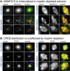
XMAP215 mislocalizes in maskin-depleted Xenopus egg extracts, but CPEB staining is unaffected. (A) Asters (left panel; 15-min time point) or spindles (right panel; 90-min time point) induced by the addition of sperm chromatin to mock-depleted (top row) or maskin-depleted (bottom rows) extract were stained with antibodies to α-tubulin (left column) and XMAP215 (middle column), as indicated on the micrographs. Right column, overlay: green, XMAP215; red, α-tubulin; blue, DNA. (B) CPEB staining of asters (left panel) or spindles (right panel) induced by addition of sperm chromatin to mock-depleted (top row) or maskin-depleted (bottom rows) extracts. Left panels, tubulin; middle panels, CPEB, right panels, overlay; green, CPEB; red, tubulin; blue, DNA. Scale bars, 25 μm.

Similar articles

Cited by

References

    1. Bellanger, J.-M. and Gönczy. P. (2003). TAC-1 and ZYG-9 form a complex that promotes microtubule assembly in C. elegans embryos. Curr. Biol. 13, 1488–1498. - PubMed
    1. Cassimeris, L., and Skibbens, R.V. (2003). Regulated assembly of the mitotic spindle: a perspective from two ends. Curr. Issues Mol. Biol. 5, 99–112. - PubMed
    1. Cassimeris, L., and Morabito, J. (2004). TOGp, the human homolog of XMAP215/Dis1, is required for centrosome integrity, spindle pole organization, and bipolar spindle assembly. Mol. Biol. Cell 15, 1580–1590. - PMC - PubMed
    1. Chen, H. M., Schmeichel, K. L., Mian, I. S., Lelievre, S., Petersen, O. W., and Bissell, M. J. (2000). AZU—A candidate breast tumor suppressor and biomarker for tumor progression. Mol. Biol. Cell. 11, 1357–1367. - PMC - PubMed
    1. Chen, X. P., Yin, H., and Huffaker., T. C. (1998). The yeast spindle pole body component Spc72p interacts with Stu2p and is required for proper microtubule assembly. J. Cell Biol. 141, 1169–1179. - PMC - PubMed

Publication types

MeSH terms

LinkOut - more resources