Comparative Study
. 2005 Mar 23;25(12):3132-41.
doi: 10.1523/JNEUROSCI.1920-04.2005.

Deleted in colorectal cancer binding netrin-1 mediates cell substrate adhesion and recruits Cdc42, Rac1, Pak1, and N-WASP into an intracellular signaling complex that promotes growth cone expansion

Affiliations
Comparative Study

Deleted in colorectal cancer binding netrin-1 mediates cell substrate adhesion and recruits Cdc42, Rac1, Pak1, and N-WASP into an intracellular signaling complex that promotes growth cone expansion

Masoud Shekarabi et al. J Neurosci. .

Abstract

Extracellular cues direct axon extension by regulating growth cone morphology. The netrin-1 receptor deleted in colorectal cancer (DCC) is required for commissural axon extension to the floor plate in the embryonic spinal cord. Here we demonstrate that challenging embryonic rat spinal commissural neurons with netrin-1, either in solution or as a substrate, causes DCC-dependent increases in growth cone surface area and filopodia number, which we term growth cone expansion. We provide evidence that DCC influences growth cone morphology by at least two mechanisms. First, DCC mediates an adhesive interaction with substrate-bound netrin-1. Second, netrin-1 binding to DCC recruits an intracellular signaling complex that directs the organization of actin. We show that netrin-1-induced growth cone expansion requires Cdc42 (cell division cycle 42), Rac1 (Ras-related C3 botulinum toxin substrate 1), Pak1 (p21-activated kinase), and N-WASP (neuronal Wiskott-Aldrich syndrome protein) and that the application of netrin-1 rapidly activates Cdc42, Rac1, and Pak1. Furthermore, netrin-1 recruits Cdc42, Rac1, Pak1, and N-WASP into a complex with the intracellular domain of DCC and Nck1. These findings suggest that DCC influences growth cone morphology by acting both as a transmembrane bridge that links extracellular netrin-1 to the actin cytoskeleton and as the core of a protein complex that directs the organization of actin.


Figures

Figure 1.
Figure 1.
Distribution of DCC in cultured embryonic rat commissural neuron growth cones. A, The dorsal halves of E13 rat spinal cords were microdissected, dissociated, and plated (D, dorsal; V, ventral; CN, commissural neurons; MN, motoneurons). B, The morphology of commissural neuron growth cones grown for 2 DIV in the presence of netrin-1 and then immunolabeled for DCC (red) and for F-actin with FITC-coupled phalloidin (green). C, DCC staining alone. D, F-actin staining alone. Scale bar, 10 μm.
Figure 2.
Figure 2.
Netrin-1 causes commissural neuron growth cone expansion. A-C, Representative examples of commissural neuron growth cone morphologies grown on a PK substrate (A) or for 5 min (B) and 30 min (C) after the addition of 80 ng/ml netrin-1 protein to the culture media. DCC immunoreactivity is shown in red. FITC-phalloidin staining of F-actin is green (100× objective). Scale bar, 10 μm. D, E, Quantification of the increase in growth cone filopodia number (D) and surface area (E) after application of 80 ng/ml netrin-1 for 5 or 30 min. Netrin-1 produced a significant increase in mean filopodia number per growth cone and growth cone surface area (*p < 0.05; **p < 0.01). Anti-DCCFB (5 μg/ml) blocked netrin-1-induced growth cone expansion (#p < 0.05; ##p < 0.01), whereas the application of anti-DCCFB alone had no effect (n = 25 per condition; error bars indicate SEM).
Figure 3.
Figure 3.
Substrate-bound netrin-1 induces growth cone expansion: evidence for an adhesive interaction between DCC and netrin-1. A-C, Commissural neuron growth cones cultured on a substrate of netrin-1 in the absence (A) or in the presence (B) of 25 μg/ml anti-netrin-1 or in the presence (C) of 5 μg/ml anti-DCC. Shown is the quantification of the number of filopodia per growth cone (D) and growth cone surface area (E) for the conditions shown in A-C. F, Quantification of cell substrate adhesion of dissociated E13 rat dorsal spinal cord cells. Representative examples of the assay are shown in G-K (10× objective lens; Hoechst-stained nuclei; grayscale inverted). A netrin-1 substrate generates a more than sevenfold increase in the number of adherent cells compared with a control BSA substrate. Preincubation of the substrates with anti-netrin-1 (I; 25 μg/ml PN3) or DCC-Fc (5 μg/ml) significantly reduced the number of adherent cells compared with netrin-1. Control IgGs (K) didnot affect adherence to netrin-1 (ANOVA; **p < 0.005 compared with control; ##p < 0.005 compared with netrin-1 or IgG control; mean ± SEM).
Figure 4.
Figure 4.
Netrin-1-induced commissural growth cone expansion requires DCC and activated Cdc42 and Rac1. A, B, Commissural neurons were infected with adenovirus expressing dominant-negative Cdc42 or Rac1 (N17Cdc42, N17Rac1), constitutively active Cdc42 or Rac1 (V12Cdc42, V12Rac1), or GFP. At 48 h after infection, 80 ng/ml netrin-1 was added, and 30 min later, the cells were fixed and immunostained. Netrin-1 significantly increased filopodia number and growth cone surface area in cells expressing GFP alone (mean ± SEM; *p < 0.05, significant increase compared with control cells expressing GFP in the absence of netrin-1). Expression of either N17Cdc42 or N17Rac1 significantly decreased the number of filopodia per growth cone and growth cone surface area (n = 25; #p < 0.05, significant decrease compared with control cells expressing GFP in the presence of netrin-1). In the absence of added netrin-1, the expression of V12Cdc42 or V12Rac1 significantly increased both filopodia number and growth cone surface area (n = 15), but to an extent significantly less than netrin-1 applied to adenoviral (Ad) GFP controls. C-E, Dissociated embryonic spinal commissural neurons were treated with 80 ng/ml netrin-1 or carrier for 5 min and then lysed and incubated with GTPγS at 31°C for 12 min. GTPγS-bound Cdc42 and Rac1 were isolated by using GST-Pak1-CRIB fusion protein and assayed by Western blotting with antibodies against Rac1 or Cdc42. Total cell lysates were probed with anti-DCC to confirm that equal amounts of total protein were loaded per lane. D, The addition of anti-DCCFB (1 or 5 μg/ml) blocked the activation of Cdc42 and Rac1 by netrin-1. E, Expression of N17Cdc42 blocked the netrin-1-dependent activation of Rac1. In contrast, expression of N17Rac1 did not block netrin-1-induced activation of Cdc42. F, Quantification of netrin-1-induced activation of endogenous Cdc42 and Rac1 in commissural neuron homogenates (after a 3 min application of 80 ng/ml netrin-1; n = 4; mean ± SEM; *p < 0.05). G, Western blot analysis of recombinant GST-Cdc42 and GST-Rac1 (∼100 ng) illustrates specificity of the antibodies against Cdc42 and Rac1. Anti-GST immunoreactivity confirms that similar amounts of recombinant protein were loaded in each lane.
Figure 5.
Figure 5.
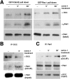
Netrin-1 promotes Cdc42, Rac1, Pak1, and DCC complex formation. A, After treatment with netrin-1 (5 or 30 min) commissural neuron cell lysates were incubated with recombinant GST-Cdc42 or GST-Rac1 and GTPγS. Phospho-Pak1 protein associated with the GST fusion proteins was isolated and analyzed by Western blot. Pak1 immunoreactivity in the whole-cell lysate (total) is shown in the last row. B, DCC was immunoprecipitated (1 μg of anti-DCCIN) from commissural neuron lysates and then analyzed by Western blot with the use of anti-Pak1. Treatment of the cells with 80 ng/ml netrin-1 for 5 min increased the amount of Pak1 protein found to coimmunoprecipitate with DCC. The coIP results are shown above Pak1 immunoreactivity in corresponding whole-cell lysates. C, Increased amounts of DCC were detected in acoIP with anti-Pak1 after treatment with 80 ng/ml netrin-1 for 5 min. The coIP results are shown above the Western blots showing immunoreactivity for Pak1 and DCC in corresponding cell lysates.
Figure 6.
Figure 6.
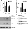
DCC-dependent activation of Pak1 by netrin-1. Commissural neurons double-immunolabeled for phospho-Pak1 (A, C) and DCC (B, D) are shown. Increased phospho-Pak1 immunoreactivity was detected in growth cones after 5 min of exposure to netrin-1 (-n in A indicates without added netrin-1). E, Quantification of phospho-Pak1 immunoreactivity (n = 28; mean ± SEM; *p < 0.05). F, G, The addition of control peptide did not affect the netrin-1-induced increase in growth cone surface area or filopodia number (mean ± SEM; *p < 0.05; ** p < 0.005). Adding the Pakpeptide 40 min before the addition of netrin-1 blocked the netrin-1-induced increase in filopodia number and growth cone surface area (n = 25; mean ± SEM; #p < 0.05; ##p < 0.005). H, The addition of 80 ng/ml netrin-1 to commissural neurons increased phospho-Pak1 as assayed by Western blot, consistent with the change in immunofluorescence shown in E. The same blot was reprobed with anti-Pak1, confirming that comparable amounts of protein are present in each lane. The netrin-1-induced increase in phospho-Pak1 was blocked by anti-DCCFB added to the cultures 1 h before the addition of netrin-1. I, Quantification of increased phospho-Pak1 as detected by Western blot analysis (after a 5 min application of 80 ng/ml netrin-1; n = 3; mean ± SEM; *p < 0.05).
Figure 7.
Figure 7.
Distribution of N-WASP and DCC in commissural neuron growth cones. Commissural neurons cultured on a control PK substrate (B, D) or 30 min after the addition of 80 ng/ml netrin-1 (A, C, E) were labeled with anti-DCC (green) and anti-N-WASP (red, A-C; white, D, E). N-WASP immunoreactivity was detected readily along the axon, throughout the growth cone, and along filopodia; C and E present an enlargement of the growth cone shown in A, illustrating the punctate distribution of N-WASP immunoreactivity. Scale bars: A, 10 μm; (in B), B-E, 5 μm.
Figure 8.
Figure 8.
N-WASP is recruited to a complex with DCC and required for netrin-1-induced growth cone expansion. A, B, After the infection with adenoviral vectors encoding either GFP or Δcof N-WASP, growth cone morphology was quantified. The addition of netrin-1 (80 ng/ml, 30 min) significantly increased the number of filopodia and growth cone surface area (*p < 0.05; **p < 0.005). Expression of Δcof N-WASP blocked the increase in both the number of filopodia (A) and surface area (B) of commissural growth cones treated with netrin-1 (#p < 0.05; ##p < 0.005). Expression of Δcof N-WASP significantly reduced the growth cone surface area below the level found in the presence of netrin-1 or control (n = 15; mean ± SEM). C, After treatment with netrin-1 (5 or 30 min) commissural neuron cell lysates were incubated with recombinant GST-Cdc42. Netrin-1 promotes the association of Pak1 and N-WASP with GST-Cdc42. Pak1 immunoreactivity in corresponding whole-cell lysates is shown in the bottom row. D, Increased amounts of DCC were detected in coIPs from commissural neuron lysates by the use of anti-N-WASP. The levels of DCC and N-WASP present in the whole-cell lysates are shown below the blot of the coIP.
Figure 9.
Figure 9.
The DCC ICD recruits a complex of signaling proteins to the plasma membrane. A, The addition of a uniform concentration of netrin-1 induces growth cone expansion, namely an increase in surface area and in the number of filopodia. B, Model of the molecular mechanisms that act downstream of netrin-1 and DCC. Nck1 binds DCC constitutively by its first and third SH3 domains. FAK, bound to the DCC ICD, recruits and activates the tyrosine kinases Src or Fyn in response to netrin-1. Netrin induces the activation of an as-yet-unidentified GEF, leading to the activation of Cdc42, Rac1, and Pak1. We speculate that the activation of a member of the Src family may regulate the activation of Cdc42 by regulating a GEF. Activated Cdc42 activates N-WASP, which promotes the nucleation of F-actin via the Arp2/3 complex.

Similar articles

Cited by

References

    1. Bagrodia S, Cerione RA (1999) Pak to the future. Trends Cell Biol 9: 350-355. - PubMed
    1. Bagrodia S, Taylor SJ, Creasy CL, Chernoff J, Cerione RA (1995) Identification of a mouse p21Cdc42/Rac activated kinase. J Biol Chem 270: 22731-22737. - PubMed
    1. Banzai Y, Miki H, Yamaguchi H, Takenawa T (2000) Essential role of neural Wiskott-Aldrich syndrome protein in neurite extension in PC12 cells and rat hippocampal primary culture cells. J Biol Chem 275: 11987-11992. - PubMed
    1. Bentley D, O'Connor TP (1994) Cytoskeletal events in growth cone steering. Curr Opin Neurobiol 4: 43-48. - PubMed
    1. Bladt F, Aippersbach E, Gelkop S, Strasser GA, Nash P, Tafuri A, Gertler FB, Pawson T (2003) The murine Nck SH2/SH3 adaptors are important for the development of mesoderm-derived embryonic structures and for regulating the cellular actin network. Mol Cell Biol 23: 4586-4597. - PMC - PubMed

Publication types

MeSH terms