

Correlative radiological, self-assessment and clinical analysis of evolution in instrumented dorsal and lateral fusion for degenerative lumbar spine disease. Autograft versus coralline hydroxyapatite
- PMID: 15789231
- PMCID: PMC3489222
- DOI: 10.1007/s00586-004-0855-5
Correlative radiological, self-assessment and clinical analysis of evolution in instrumented dorsal and lateral fusion for degenerative lumbar spine disease. Autograft versus coralline hydroxyapatite
Abstract
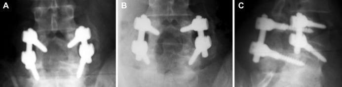
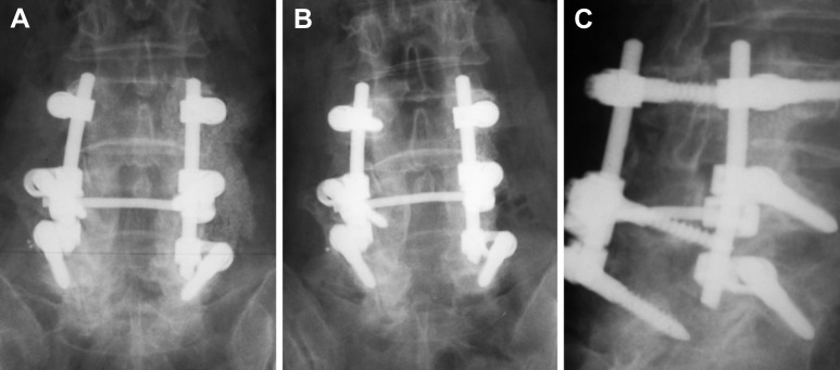
This prospective longitudinal randomized clinical and radiological study compared the evolution of instrumented posterolateral lumbar and lumbosacral fusion using either coralline hydroxyapatite (CH), or iliac bone graft (IBG) or both in three comparable groups, A, B and C, which included 19, 18 and 20 patients, respectively, who suffered from symptomatic degenerative lumbar spinal stenosis and underwent decompression and fusion. The patients were divided randomly according to the graft used and the side that it was applied. The spines of group A received autologous IBG bilaterally; group B, IBG on the left side and hydroxyapatite mixed with local bone and bone marrow on the right side; group C, hydroxyapatite mixed with local bone and bone marrow bilaterally. The age of the patients in the groups A, B and C was 61+/-11 years, 64+/-8 years and 58+/-8 years, respectively. The SF-36, Oswestry Disability Index (ODI), and Roland-Morris (R-M) surveys were used for subjective evaluation of the result of the surgery and the Visual Analogue Scale (VAS) for pain severity. Plain roentgenograms including anteroposterior, lateral and oblique views, and lateral plus frontal bending views of the instrumented spine and CT scan were used to evaluate the evolution of the posterolateral fusion in all groups and sides. Two independent senior orthopaedic radiologists were asked to evaluate first the evolution of the dorsolateral bony fusion 3-48 months postoperatively with the Christiansen's radiologic method, and secondly the hydroxyapatite resorption course in the spines of groups B and C. The diagnosis of solid spinal fusion was definitively confirmed with the addition of the bending views, CT scans and self-assessment scores. The intraobserver and interobserver agreement (r) for radiological fusion was 0.71 and 0.69, respectively, and 0.83 and 0.76 for evaluation of CH resorption. T(12)-S(1) lordosis and segmental angulation did not change postoperatively. There was no radiological evidence for non-union on the plain roentgenograms and CT scans. Radiological fusion was achieved 1 year postoperatively and was observed in all groups and vertebral segments. Six months postoperatively there was an obvious resorption of hydroxyapatite granules at the intertransverse intersegmental spaces in the right side of the spines of group B and both sides of group C. The resorption of hydroxyapatite was completed 1 year postoperatively. Bone bridging started in the third month postoperatively in all instrumented spines and all levels posteriorly as well as between the transverse processes in the spines of the group A and on the left side of the spines of group B where IBG was applied. SF-36, ODI, and R-M score improved postoperatively in a similar way in all groups. There was one pedicle screw breakage at the lowermost instrumented level in group A and two in group C without radiologically visible pseudarthrosis, which were considered as having non-union. Operative time and blood loss were less in the patients of group C, while donor site complaints were observed in the patients of the groups A and B only. This study showed that autologous IBG remains the "gold standard" for achieving solid posterior instrumented lumbar fusion, to which each new graft should be compared. The incorporation of coralline hydroxyapatite mixed with local bone and bone marrow needs adequate bleeding bone surface. Subsequently, hydroxyapatite was proven in this series to not be appropriate for intertransverse posterolateral fusion, because the host bone in this area is little. However, the use of hydroxyapatite over the decorticated laminae that represents a wide host area was followed by solid dorsal fusion within the expected time.
Figures

Similar articles
-
Rigid, semirigid versus dynamic instrumentation for degenerative lumbar spinal stenosis: a correlative radiological and clinical analysis of short-term results.Spine (Phila Pa 1976). 2004 Apr 1;29(7):735-42. doi: 10.1097/01.brs.0000112072.83196.0f. Spine (Phila Pa 1976). 2004. PMID: 15087795 Clinical Trial.
-
Does Wallis implant reduce adjacent segment degeneration above lumbosacral instrumented fusion?Eur Spine J. 2009 Jun;18(6):830-40. doi: 10.1007/s00586-009-0976-y. Epub 2009 Apr 23. Eur Spine J. 2009. PMID: 19387697 Free PMC article. Clinical Trial.
-
Single-level instrumented posterolateral fusion of lumbar spine with beta-tricalcium phosphate versus autograft: a prospective, randomized study with 3-year follow-up.Spine (Phila Pa 1976). 2008 May 20;33(12):1299-304. doi: 10.1097/BRS.0b013e3181732a8e. Spine (Phila Pa 1976). 2008. PMID: 18496340 Clinical Trial.
-
In vivo evaluation of coralline hydroxyapatite and direct current electrical stimulation in lumbar spinal fusion.Spine (Phila Pa 1976). 1999 Oct 15;24(20):2127-33. doi: 10.1097/00007632-199910150-00012. Spine (Phila Pa 1976). 1999. PMID: 10543011
-
Results of lumbar spondylodeses using different bone grafting materials after transforaminal lumbar interbody fusion (TLIF).Eur Spine J. 2017 Nov;26(11):2835-2842. doi: 10.1007/s00586-017-5145-0. Epub 2017 May 25. Eur Spine J. 2017. PMID: 28547573 Clinical Trial.
Cited by
-
Bone Substitute Options for Spine Fusion in Patients With Spine Trauma-Part I: Fusion Biology, Autografts, Allografts, Demineralized Bone Matrix, and Ceramics.Korean J Neurotrauma. 2023 Dec 19;19(4):446-453. doi: 10.13004/kjnt.2023.19.e62. eCollection 2023 Dec. Korean J Neurotrauma. 2023. PMID: 38222832 Free PMC article. Review.
-
[Bone substitutes in scoliosis surgery].Orthopade. 2009 Feb;38(2):181-8. doi: 10.1007/s00132-008-1369-3. Orthopade. 2009. PMID: 19093096 Review. German.
-
Bone graft materials for posterolateral fusion made simple: a systematic review.Eur Spine J. 2018 Aug;27(8):1856-1867. doi: 10.1007/s00586-018-5511-6. Epub 2018 Feb 14. Eur Spine J. 2018. PMID: 29445947
-
Bone substitutes and expanders in Spine Surgery: A review of their fusion efficacies.Int J Spine Surg. 2016 Sep 22;10:33. doi: 10.14444/3033. eCollection 2016. Int J Spine Surg. 2016. PMID: 27909654 Free PMC article.
-
Ceramic Biologics for Bony Fusion-a Journey from First to Third Generations.Curr Rev Musculoskelet Med. 2020 Aug;13(4):530-536. doi: 10.1007/s12178-020-09651-x. Curr Rev Musculoskelet Med. 2020. PMID: 32562147 Free PMC article. Review.
References
-
- Abott LC (1944) The use of iliac bone in the treatment of ununited fractures. In: Instructional Course Lectures. The American Academy of Orthopaedic Surgeons, Park Ridge, pp 13–22
-
- An HS, Simpson JM, Glover JM, Stephany J. Comparison between allograft plus demineralized bone matrix versus autograft in anterior cervical fusion: a prospective multicenter study. Spine. 1995;20:2211–2216. - PubMed
-
- Banwart JA, Asher MA, Hassanein RS. Iliac crest bone graft harvest donor site morbidity. A statistical evaluation. Spine. 1995;20:1055–1060. - PubMed
-
- Baramki GH, Steffen T, Lander P, Chang M, Marchesi D. The efficacy of interconnected porous hydroxyapatite in achieving posterolateral lumbar fusion in sheep. Spine. 2000;25:1053–1060. - PubMed
Publication types
MeSH terms
Substances
LinkOut - more resources
Full Text Sources
Medical
