. 2005 Apr 5;102(14):5227-32.
doi: 10.1073/pnas.0501029102. Epub 2005 Mar 24.

Adult human hematopoietic stem cells produce neurons efficiently in the regenerating chicken embryo spinal cord

Affiliations

Adult human hematopoietic stem cells produce neurons efficiently in the regenerating chicken embryo spinal cord

Olafur E Sigurjonsson et al. Proc Natl Acad Sci U S A. .

Abstract

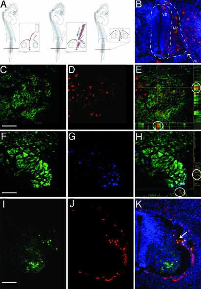
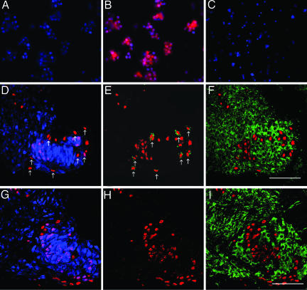
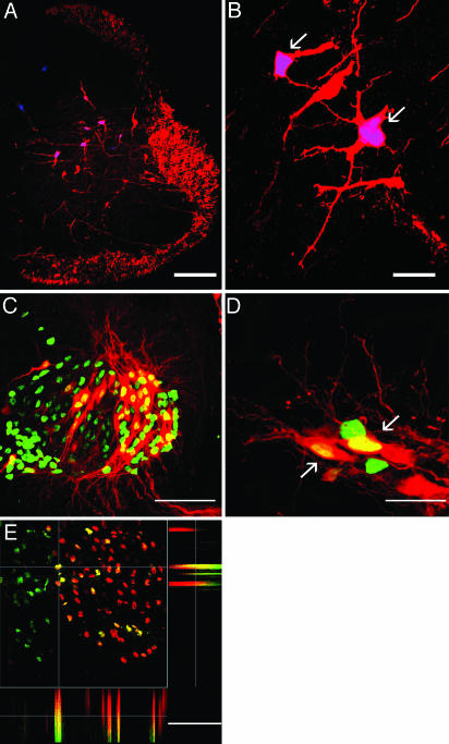
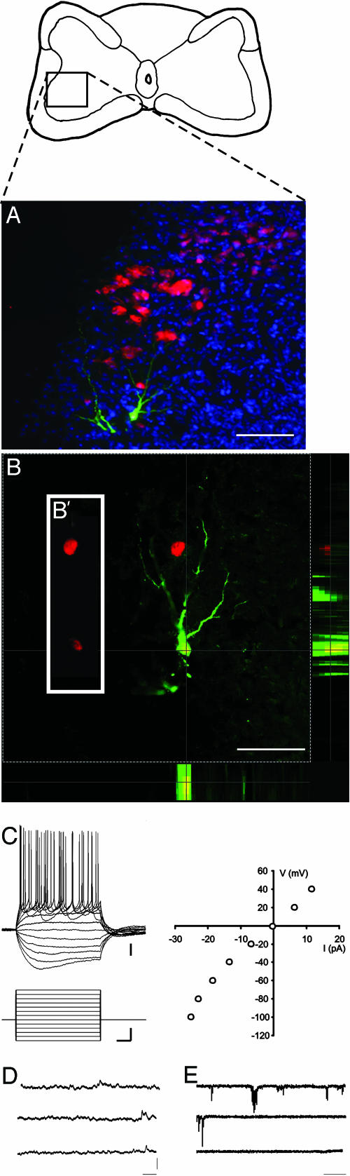
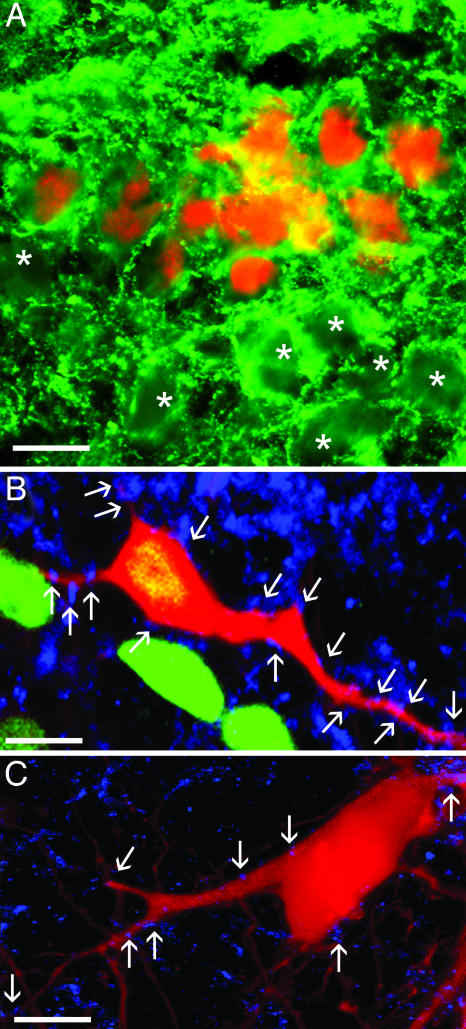
Hematopoietic stem cells (HSCs) have been proposed as a potential source of neural cells for use in repairing brain lesions, but previous studies indicate a low rate of neuronal differentiation and have not provided definite evidence of neuronal phenotype. To test the neurogenic potential of human HSCs, we implanted CD34+ HSCs from adult human bone marrow into lesions of the developing spinal cord in the chicken embryo and followed their differentiation by using immunohistochemistry, retrograde labeling, and electrophysiology. We find that human cells derived from the implanted population express the neuronal markers NeuN and MAP2 at substantially higher rates than previously reported. We also find that these cells exhibit neuronal cytoarchitecture, extend axons into the ventral roots or several segments in length within the spinal white matter, are decorated with synaptotagmin+ and GABA+ synaptic terminals, and exhibit active membrane properties and spontaneous synaptic potentials characteristic of functionally integrated neurons. Neuronal differentiation is accompanied by loss of CD34 expression. Careful examination with confocal microscopy reveals no signs of heterokaryons, and human cells never express a chicken-specific antigen, suggesting that fusion with host chicken cells is unlikely. We conclude that the microenvironment in the regenerating spinal cord of the chicken embryo stimulates substantial proportions of adult human HSCs to differentiate into full-fledged neurons. This may open new possibilities for a high-yield production of neurons from a patient's own bone marrow.


Figures

Fig. 1.
Fig. 1.
Expression of neuronal markers by human cells after integration into the regenerated embryonic spinal cord. (A) A one- to three-segment-long portion of the lumbar neural tube was excised unilaterally, and hHSCs were injected into the lesion with a micropipette. (B) PKH26-labeled (red) human cells integrated into the regenerated spinal cord. Nuclei counterstained blue with Hoechst 33342. (C–E) MAP2-(green) and hNA-(red) labeled human cells in ventral horn; colocalization demonstrated (circles) for one selected cell (at cross hairs) shown in side views of confocal stack (E). (F–H) NeuN-(green) and hNA-labeled (blue) human cells in ventral horn; colocalization demonstrated (circles) for one selected cell (at cross hairs) shown in side views of confocal stack (H). (I–K) Human cells (red, hNA) implanted outside the developing spinal cord do not invade neural tissue or express neuronal antigens (MAP2, green) except at small lesion site (arrow). Nuclei counterstained with Hoechst 33342 (blue). VZ, ventricular zone; MZ, mantle zone; VH, ventral horn. All images are from 10-μm transverse sections, dorsal up; B–H are of 0.5-μm optical sections. [Bars, 60 (B), 40 (C–H), and 80 (I–K) μm.]
Fig. 2.
Fig. 2.
Neuronal differentiation by human cells involves loss of CD34 expression but never expression of a chicken-specific marker. (A–C) Freshly isolated CD34+ cells (B, red) do not express NeuN (A) or MAP2 (C). Nuclei counterstained with Hoechst 33342 (blue). (D–I) Expression of NeuN (blue), hNA (red), CD34 (green, E and H), and chicken-specific marker (green; F and I)3(D–F) and 5 days (G–I) after implantation of hHSCs. Arrows point to hNA+ cells that are NeuN-negative (D) and CD34-positive (E). F and I show the sections in D and G superimposed on neighboring sections. A–C are images of cytospins, and D–I are 0.5-μm optical sections from 10-μm transverse sections, dorsal up. [Bars, 40 μm(D–I).]
Fig. 3.
Fig. 3.
Neuronal morphology and expression of motoneuron-specific markers by human cells in the regenerated embryonic spinal cord. (A and B) Human cells retrogradely labeled from the ventral funiculus [arrows; red, rhodamine dextran amine (RDA); blue, hNA] are indistinguishable from spinal interneurons. (C and D) Human cells retrogradely labeled from peripheral nerve (arrows; red, RDA; green, hNA). (E) Colocalization of Islet-1 (red) and hNA (green), demonstrated for one selected cell (at cross hairs) in side views of confocal stack. A, B, and E are 0.5-μm optical sections, and C and D are confocal stacks from 10-μm transverse sections, dorsal up. [Bars, 150 (A), 15 (B), 40 (C), 10 (D), and 40 (E) μm.]
Fig. 4.
Fig. 4.
Electrophysiological properties of human neurons. (A) Transverse section through spinal cord 10 days after implantation of human cells (red, hNA), with one human cell labeled intracellularly with biocytin (green), counterstained with Hoechst 33324 (blue). Diagram above shows placement of the field shown within the cross-sectional area of the cord. (B) Optical section through this neuron, showing colocalization of biocytin (green) and hNA (red) in side views of confocal stack. B′ is a slice of the single optical section, showing the red channel only, displaced to the left to emphasize that the biocytin-labeled neuron expresses hNA. (C) Current steps (600 ms, incrementing + 20 from –120 pA) in current–clamp mode show that the human cell is excitable, fires overshooting action potentials in response to depolarization, and exhibits a voltage sag in response to hyperpolarizing pulses. I–V curve shown to the right. (D and E) Spontaneous synaptic events observed at resting membrane potential (–52 mV) in current–(D) and voltage–clamp (E) mode. The cell had an input resistance of 375 megaohms and a membrane time constant of 45 ms. [Bars, 100 μm(A); 60 μm(B); 10 mV, 60 pA, and 100 ms (C); 5 mV(D); 50 pA (E); and 1 sec (D and E).
Fig. 5.
Fig. 5.
Morphological evidence of synaptic contacts onto human cells. (A) Human cells (red, hNA) are decorated with GABA+ synaptic terminals (green), as are nearby chicken neurons (asterisks). (B) Human (green, hNA) and (C) chicken neurons, retrogradely labeled from peripheral nerve (red, RDA), decorated with synaptotagmin+ terminals (blue). A–C are from 10-μm transverse sections, dorsal up. B and C are 0.5-μm optical sections. [Bars, 20 μm(A) and 10 μm(B and C).]

References

    1. Weimann, J. M., Charlton, C. A., Brazelton, T. R., Hackman, R. C. & Blau, H. M. (2003) Proc. Natl. Acad. Sci. USA 100, 2088–2093. - PMC - PubMed
    1. Sanchez-Ramos, J., Song, S., Cardozo-Pelaez, F., Hazzi, C., Stedeford, T., Willing, A., Freeman, T. B., Saporta, S., Janssen, W., Patel, N., et al. (2000) Exp. Neurol. 164, 247–256. - PubMed
    1. Mezey, E., Key, S., Vogelsang, G., Szalayova, I., Lange, G. D. & Crain, B. (2003) Proc. Natl. Acad. Sci. USA 100, 1364–1369. - PMC - PubMed
    1. Mezey, E., Chandross, K. J., Harta, G., Maki, R. A. & McKercher, S. R. (2000) Science 290, 1779–1782. - PubMed
    1. Song, H. J., Stevens, C. F. & Gage, F. H. (2002) Nat. Neurosci. 5, 438–445. - PubMed

Publication types

MeSH terms