. 2005 Apr 5;102(14):5138-43.
doi: 10.1073/pnas.0501675102. Epub 2005 Mar 24.

Foxp3 interacts with nuclear factor of activated T cells and NF-kappa B to repress cytokine gene expression and effector functions of T helper cells

Affiliations

Foxp3 interacts with nuclear factor of activated T cells and NF-kappa B to repress cytokine gene expression and effector functions of T helper cells

Estelle Bettelli et al. Proc Natl Acad Sci U S A. .

Abstract

Scurfy mice, which are deficient in a functional Foxp3, exhibit a severe lymphoproliferative disorder and display generalized over-production of cytokines. Here, we show that, among the Foxp transcriptional factor family, which includes Foxp1, Foxp2, and Foxp3, only Foxp3 has the ability to inhibit IL-2, IL-4, and IFN-gamma production by primary T helper cells. We found that Foxp3 physically associates with the Rel family transcription factors, nuclear factor of activated T cells (NFAT) and NF-kappaB, and blocks their ability to induce the endogenous expression of their target genes, including key cytokine genes. More importantly, T cells derived from scurfy mice have a dramatic increase in nuclear factor of activated T cells (NFAT) and NF-kappa B transcriptional activity compared with the T cells derived from WT mice. Furthermore, complementation of Foxp3 in scurfy-derived T cells lowers the NFAT and NF-kappa B transcriptional activity to the physiological level. Finally, we show that myelin proteolipid protein-specific autoreactive T cells transduced with Foxp3 cannot mediate experimental autoimmune encephalomyelitis, providing further support that Foxp3 suppresses the effector function of autoreactive T cells. Foxp3 has already been associated with the generation of CD4(+)CD25+ regulatory T cells; our data additionally demonstrate that Foxp3 suppresses the effector functions of T helper cells by directly inhibiting the activity of two key transcription factors, NFAT and NF-kappa B, which are essential for cytokine gene expression and T cell functions.


Figures
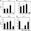
Fig. 1.
Fig. 1.
Foxp3, but not other Foxp family members, suppresses cytokine expression in T cells. Foxp1, Foxp2, Foxp3, or deletion mutants of Foxp3 cDNAs (encoding the indicated amino acids) were inserted into the RV GFP retrovirus vector. These vectors simultaneously express a specific Foxp cDNA with the GFP, with the use of an internal ribosomal entry site (IRES). Purified T cells were stimulated with anti-CD3 and anti-CD28, and infected with the Foxp-expressing or control retroviruses in the presence of human IL-2. The GFP+ T cells were sorted 48 h postinfection. One week after the infection, GFP+-infected CD4+ T cells were stimulated with antigen-presenting cells (irradiated splenocytes) plus soluble anti-CD3 antibody. The figure represents the amount of IL-2 (A and D), IL-4 (B), and IFN-γ (C) secreted in the culture supernatant of these activated cells, as measured by ELISA.
Fig. 2.
Fig. 2.
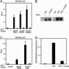
Foxp3 inhibits NF-κB transcriptional activity and represses the endogenous expression of the NF-κB target gene, A20. (A and C) 293T cells were transfected with the indicated plasmids along with NF-κB luciferase reporter and Tk-Renilla reporter as an internal control. Twenty-four hours later (and an additional 6 h when cells were stimulated with TNF-α), cells were analyzed by means of the dual luciferase assay, with relative activity determined after normalization for Renilla.(B) 293T cells were transfected with Foxp3 and 48 h later stimulated for 1 h with TNF-α or transfected with P65 along with Foxp3, and NF-κB DNA-binding activity was assessed by EMSA. (D) 293T cells were transfected with the indicated plasmids, and, 48 h posttransfection, endogenous A20 expression was determined by real-time PCR.
Fig. 3.
Fig. 3.
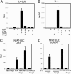
Foxp3 inhibits NFAT transcriptional activity and represses the production of endogenous IL-4. (A) M12 cells were transiently transfected with the indicated expression plasmids along with the IL-4 luciferase reporter. After transfection, half of the transfected cells were cultured for 24 h and assayed for luciferase activity. (B) Cells from the other half were cultured for 72 h, and the supernatants were assayed for IL-4. Results are representative of three independent experiments. 293T cells (C) or Jurkat cells (D) were transfected with NFATp, a constitutively active NFATp (NFAT-CA), or a pcDNA3 vector control (Vec.) along with or without Foxp3 as indicated. In all cases, the NFAT luciferase reporter and the TK-Renilla reporter (as an internal control) were used (see Materials and Methods). Twenty-four hours later, cells were analyzed by means of the dual luciferase assay, with relative activity determined after normalization with Renilla. (D) Jurkat cells were transiently transfected as indicated above and analyzed by means of the dual luciferase assay after 6 h of stimulation with phorbol 12-myristate 13-acetate (PMA) and ionomycin (P/I).
Fig. 4.
Fig. 4.
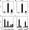
Physical association between Foxp3 and the REL proteins NFATp and NF-κB and repression of their transactivation domain. (A) 293T cells were transiently transfected with P65, or with P65-GFP, along with Myc-Foxp3 or control vector and immunoprecipitated by using an anti-Myc monoclonal antibody. Proteins were run on SDS/PAGE and immunoblotted with a rabbit anti-P65 polyclonal antibody to detect the immunoprecipitates, and with anti-p65 or anti-Myc antibodies for the lysates. (B) 293T cells were transduced with RV control or with RV-HA-Foxp3, stimulated with TNF-α for 1 h, and immunoprecipitated by using an anti-HA monoclonal antibody and immunoblotted with a rabbit anti-P65 polyclonal antibody to detect the immunoprecipitate and the lysates. (C) 293T cells were transiently transfected with HA-tagged NFAT-CA or RSK2 along with Myc-Foxp3 or control vector and immunoprecipitated by using an anti-Myc monoclonal antibody. The immunoblotting was performed with a rabbit anti-HA polyclonal antibody to detect the immunoprecipitates and the lysates. (D) 293T cells were transfected with a Gal4 luciferase reporter along with a Gal4 DNA-binding domain, Gal4-P65, Gal4-NFAT, Gal4-KRC, or Gal4-ELK1 with or without Foxp3. Twenty-four hours later, cells were harvested and analyzed for luciferase activity as described in Materials and Methods.
Fig. 5.
Fig. 5.
The absence of a functional Foxp3 leads to NF-κB and NFAT hyper-activation. NFAT (A) and NF-κB (B) luciferase activities were assessed in primary CD4+ T cells derived from WT scurfy mice (Scurf.) or scurfy T cells transfected with Foxp3 and activated with anti-CD3 and anti-CD28 for 24 h. Primary T cells derived from WT (C) or from scurfy mice (D) were transfected with a Gal4 luciferase reporter along with a Gal4 DNA-binding domain, Gal4-P65, Gal4-NFAT, or Gal4-ELK1 with or without Foxp3 and activated with anti-CD3 and anti-CD28. Twenty-four hours later, cells were harvested and analyzed for luciferase activity as described in Materials and Methods.
Fig. 6.
Fig. 6.
Effect of Foxp3 on the in vivo effector function of CD4+ T cells. (A) CD4+ T cells from PLP-specific TCR transgenic mice were purified, activated with PLP139-151, and infected with RV or RV-Foxp3 retroviruses. CD4+ GFP+ from RV- or RV-Foxp3-infected cells were then sorted and tested for their proliferative response toward PLP peptide by 3H incorporation. The data represent the proliferative response ± SE. (B) The figure represents the amount of IL-2 and IFN-γ secreted in the culture supernatant of these activated cells as measured by ELISA. (C) GFP+-sorted PLP-specific T cells (5 × 10⁶) infected with RV or RV-Foxp3 were injected into RAG-deficient mice. The progression of EAE was followed over time and is presented as the mean clinical disease among each group.

References

    1. Kaufmann, E. & Knochel, W. (1996) Mech. Dev. 57, 3–20. - PubMed
    1. Carlsson, P. & Mahlapuu, M. (2002) Dev. Biol. 250, 1–23. - PubMed
    1. Banerjee-Basu, S. & Baxevanis, A. D. (2004) Proteins 54, 639–647. - PubMed
    1. Banham, A. H., Beasley, N., Campo, E., Fernandez, P. L., Fidler, C., Gatter, K., Jones, M., Mason, D. Y., Prime, J. E., Trougouboff, P., et al. (2001) Cancer Res. 61, 8820–8829. - PubMed
    1. Barrans, S. L., Fenton, J. A., Banham, A., Owen, R. G. & Jack, A. S. (2004) Blood 104, 2933–2935. - PubMed

Publication types

MeSH terms