. 2005 Apr 20;24(8):1523-36.
doi: 10.1038/sj.emboj.7600632. Epub 2005 Mar 24.

Structure of P-protein of the glycine cleavage system: implications for nonketotic hyperglycinemia

Affiliations

Structure of P-protein of the glycine cleavage system: implications for nonketotic hyperglycinemia

Tadashi Nakai et al. EMBO J. .

Abstract

The crystal structure of the P-protein of the glycine cleavage system from Thermus thermophilus HB8 has been determined. This is the first reported crystal structure of a P-protein, and it reveals that P-proteins do not involve the alpha(2)-type active dimer universally observed in the evolutionarily related pyridoxal 5'-phosphate (PLP)-dependent enzymes. Instead, novel alphabeta-type dimers associate to form an alpha(2)beta(2) tetramer, where the alpha- and beta-subunits are structurally similar and appear to have arisen by gene duplication and subsequent divergence with a loss of one active site. The binding of PLP to the apoenzyme induces large open-closed conformational changes, with residues moving up to 13.5 A. The structure of the complex formed by the holoenzyme bound to an inhibitor, (aminooxy)acetate, suggests residues that may be responsible for substrate recognition. The molecular surface around the lipoamide-binding channel shows conservation of positively charged residues, which are possibly involved in complex formation with the H-protein. These results provide insights into the molecular basis of nonketotic hyperglycinemia.


Figures

Figure 1
Figure 1
The multistep reaction catalyzed by the glycine cleavage system. First, P-protein catalyzes the decarboxylation of the glycine molecule concomitantly with the transfer of the residual aminomethyl group to a sulfur atom on the lipoyl group of the oxidized H-protein (Hox), generating the aminomethylated H-protein (Ham). Next, the T-protein catalyzes the transfer of a methylene group from Ham to tetrahydrofolate (THF), resulting in the release of NH3 and the generation of reduced H-protein (Hred). Finally, the dihydrolipoyl group of Hred is oxidized by L-protein and the lipoyl group of Hox is regenerated, thereby completing the catalytic cycle. The figure is adapted from Douce et al (2001).
Figure 2
Figure 2
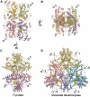
Overall structures of P-protein and GluDC, the closest homolog with a known structure. Front view (A), top view (B) and side view (C) of (αNβC)2-tetrameric P-protein, and side view of αI6 hexameric GluDC (D). PLP molecules are represented by yellow spheres.
Figure 3
Figure 3
Dimer and subunit structures of P-protein and GluDC. Stereoview of the αNβC dimer of the P-protein (A) and view of αI2 dimer of GluDC (B). Stereoviews of αN (C) and βC (D) of the P-protein. Superposition of αN (green) and βC (orange) of the P-protein, and αI of GluDC (purple) (E). PLP molecules and their bound lysine residues are shown as ball-and-stick models. In panels C and D, topology diagrams are included, in which α-helices and β-strands are represented by circles and triangles, respectively. The diagrams were generated by Tops (Westhead et al, 1999) with manual modifications.
Figure 4
Figure 4
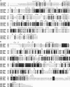
Sequence alignment of P-proteins with secondary structure assignment of Tth P-protein: (A) αN and (B) βC. The amino-acid sequences were obtained from the Swiss-Prot database. The multiple alignments of these sequences with those of Tth P-protein (GCSA_THETH and GCSB_THETH) were performed using the program CLUSTAL W (Thompson et al, 1994). The figure was produced using ESPRIPT (Gouet et al, 1999). The sequence names correspond to the Swiss-Prot accession IDs, except for the Tth P-protein. The residue numbering of the human P-protein starts from 40 since the N-terminal presequence is omitted. The numbering of βC of Tth P-protein starts from 439, as if αN and βC consisted of a single polypeptide, in order to facilitate comparisons between the αNC2-dimeric and (αNβC)2-tetrameric P-proteins.
Figure 5
Figure 5
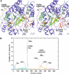
Conformational changes upon binding of the cofactor PLP. (A) Stereoview of the superimposition of the Pholo and Papo (semitransparent) structures by least-squares fitting of Cα atoms in immobile regions (see text), where the displacements are represented by a color gradient from blue (<0.37 Å) to red (>2.50 Å). The orientation shows the protein rotated by 180° around a vertical axis with respect to Figure 3A. (B) Displacements for equivalent Cα atoms between the two structures after the superimposition. The curves are colored by domain according to Figure 3A, except for the black curve indicating the displacements after the superimposition of only the mobile subdomains, in which the values are shifted by +6.0 Å unit in order to improve clarity.
Figure 6
Figure 6
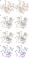
Structure of the active-site pocket of P-protein. Stereoviews of the cofactor-binding site for Pholo (A); the substrate-binding site for Pholo (B) and for Pholo·AOA (C); and superposition of the corresponding sites between Pholo (cyan) and Pholo·AOA (magenta) (D). Panels B and C include the FoFc simulated-annealing omit map contoured at 3.5σ around Lys704β, PLP and AOA. After removing them and their neighboring residues, a simulated-annealing map was calculated with a starting temperature of 1000 K. In panels A–C, carbon atoms of each subunit are colored according to Figure 2, whereas the α and β designations in residue labels are omitted. Water molecules are shown as red spheres. Dashed lines represent hydrogen bonds.
Figure 7
Figure 7
Lipoamide-binding channel and the molecular surface properties neighboring its entrance. (A) Ribbon diagrams with molecular surfaces of the P-protein (upper) and the H-protein (lower), where PLP and the lipoyl-lysine arm are represented by yellow and cyan spheres, respectively. (B) Electrostatic potential surfaces, onto which the negative (red) and positive (blue) charges are mapped. (C) Space-filling models, colored as follows: red, fully conserved residues; yellow, conservation of ‘strong' group residues; green, conservation of ‘weaker' group residues, where the groups are according to the CLUSTAL W (Thompson et al, 1994) manual. (D) Lipoamide-binding channel viewed from its entrance: molecular surface (left) and the residues constituting the channel (right, stereoview). The model structure for the lipoyl-lysine arm was manually docked into the channel: first, the aminomethylated lipoyl-lysine was connected to PLP via an aldimine bond, as shown in Supplementary Figure 1E, in place of AOA and positioned within the channel, avoiding significant steric clashes; then, the aminomethyl group was removed.
Figure 8
Figure 8
Stereoview of the NKH mutations mapped on the model structure of the human P-protein. The 11 point mutations associated with the disease are shown in magenta. The orientation is the same as that in Figure 3A. The linker region connecting αNC-N and αNC-C is represented by a cyan curve with spheres placed at intervals of 3.8 Å (the mean length between two successive Cα atoms), while the corresponding region of Tth P-protein is shown in semitransparent gray. The other regions are colored by domain according to Figure 3A. In constructing the model, we started with the Tth P-protein backbone, and then added a linker region along the molecular surface, so as to select the shortest distance between the αN C-terminus (Leu437α in Tth) and one of the residues in the N-terminal region of βC or βC′, assuming that the overall arrangement of the human αNC2 dimer subunits is similar to that of the TthNβC)2 tetramer. The resulting linker region starts from Leu437α and leads to Leu467β, and is composed of 13 residues. Our model linker is shorter than the actual linker in the human P-protein, which consists of 17 residues connecting Ser492 with Ile508. The human P-protein linker thus may contain helical and other regions that are similar to the corresponding region in the Tth P-protein.

References

    1. Adachi S, Oguchi T, Tanida H, Park SY, Shimizu H, Miyatake H, Kamiya N, Shiro Y, Inoue Y, Ueki T, Iizuka T (2001) The RIKEN structural biology beamline II (BL44B2) at the SPring-8. Nucl Instrum Methods A 467: 711–714
    1. Alexeev D, Alexeeva M, Baxter RL, Campopiano DJ, Webster SP, Sawyer L (1998) The crystal structure of 8-amino-7-oxononanoate synthase: a bacterial PLP-dependent, acyl-CoA-condensing enzyme. J Mol Biol 284: 401–419 - PubMed
    1. Applegarth DA, Toone JR (2001) Nonketotic hyperglycinemia (glycine encephalopathy): laboratory diagnosis. Mol Genet Metab 74: 139–146 - PubMed
    1. Bertoldi M, Gonsalvi M, Contestabile R, Voltattorni CB (2002) Mutation of tyrosine 332 to phenylalanine converts dopa decarboxylase into a decarboxylation-dependent oxidative deaminase. J Biol Chem 277: 36357–36362 - PubMed
    1. Bourguignon J, Neuburger M, Douce R (1988) Resolution and characterization of the glycine-cleavage reaction in pea leaf mitochondria. Properties of the forward reaction catalysed by glycine decarboxylase and serine hydroxymethyltransferase. Biochem J 255: 169–178 - PMC - PubMed

Publication types

MeSH terms