

Coronary flow reserve in stress-echo lab. From pathophysiologic toy to diagnostic tool
- PMID: 15792499
- PMCID: PMC1084250
- DOI: 10.1186/1476-7120-3-8
Coronary flow reserve in stress-echo lab. From pathophysiologic toy to diagnostic tool
Abstract
The assessment of coronary flow reserve by transthoracic echocardiography has recently been introduced into clinical practice with gratifying results for the diagnosis of left anterior descending artery disease simultaneously reported by several independent laboratories. This technological novelty is changing the practice of stress echo for 3 main reasons. First, adding coronary flow reserve to regional wall motion allows us to have - in the same sitting - high specificity (regional wall motion) and a high sensitivity (coronary flow reserve) diagnostic marker, with an obvious improvement in overall diagnostic accuracy. Second, the technicalities of coronary flow reserve shift the balance of stress choice in favour of vasodilators, which are a more robust hyperemic stress and are substantially easier to perform with dual imaging than dobutamine or exercise. Third, the coronary flow reserve adds a quantitative support to the exquisitely qualitative assessment of wall motion analysis, thereby facilitating the communication of stress echo results to the cardiological world outside the echo lab. The next challenges involve the need to expand the exploration of coronary flow reserve to the right and circumflex coronary artery and to prove the additional prognostic value - if any - of coronary flow reserve over regional wall motion analysis, which remains the cornerstone of clinically-driven diagnosis in the stress echo lab.
Figures

References
-
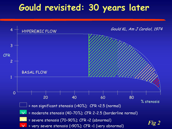
- White CW, Wright CB, Doty DB, Hiratzka LF, Eastham CL, Harrison DG, Marcus ML. Does visual interpretation of the coronary arteriogram predict the physiologic importance of a coronary stenosis? N Engl J Med. 1984;310:819–824. - PubMed
-
- Topol EJ, Nissen SE. Our preoccupation with coronary luminology. The dissociation between clinical and angiographic findings in ischemic heart disease. Circulation. 1995;92:2333–42. - PubMed
-
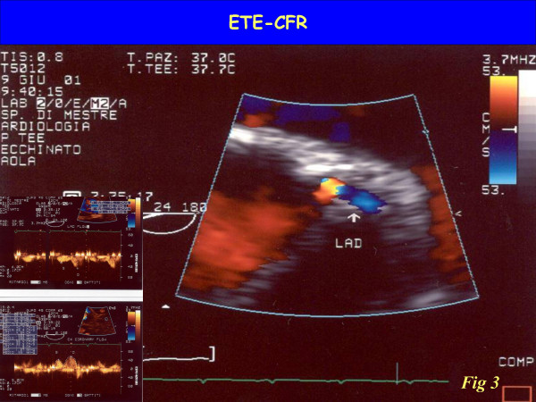
- Iliceto S, Marangelli V, Memmola C, Rizzon P. Transesophageal Doppler echocardiography evaluation of coronary blood flow velocity in baseline conditions and during dipyridamole-induced coronary vasodilation. Circulation. 1991;83:61–9. - PubMed
Publication types
MeSH terms
LinkOut - more resources
Full Text Sources
Medical
Miscellaneous
