. 2005 Apr 5;102(14):5144-9.
doi: 10.1073/pnas.0501463102. Epub 2005 Mar 25.

Varicelloviruses avoid T cell recognition by UL49.5-mediated inactivation of the transporter associated with antigen processing

Affiliations

Varicelloviruses avoid T cell recognition by UL49.5-mediated inactivation of the transporter associated with antigen processing

Danijela Koppers-Lalic et al. Proc Natl Acad Sci U S A. .

Abstract

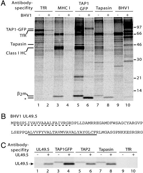
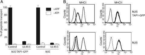
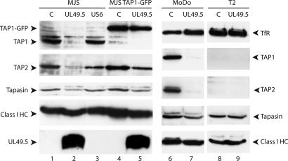
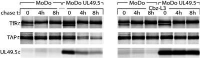
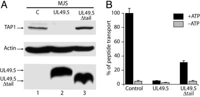
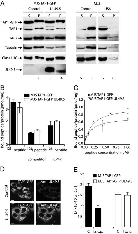
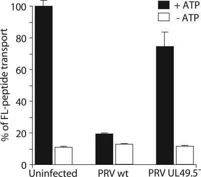
Detection and elimination of virus-infected cells by cytotoxic T lymphocytes depends on recognition of virus-derived peptides presented by MHC class I molecules. A critical step in this process is the translocation of peptides from the cytoplasm into the endoplasmic reticulum by the transporter associated with antigen processing (TAP). Here, we identified the bovine herpesvirus 1-encoded UL49.5 protein as a potent inhibitor of TAP. The expression of UL49.5 results in down-regulation of MHC class I molecules at the cell surface and inhibits detection and lysis of the cells by cytotoxic T lymphocytes. UL49.5 homologs encoded by two other varicelloviruses, pseudorabies-virus and equine herpesvirus 1, also block TAP. Homologs of UL49.5 are widely present in herpesviruses, acting as interaction partners for glycoprotein M, but in several varicelloviruses UL49.5 has uniquely evolved additional functions that mediate its participation in TAP inhibition. Inactivation of TAP by UL49.5 involves two events: inhibition of peptide transport through a conformational arrest of the transporter and degradation of TAP by proteasomes. UL49.5 is degraded along with TAP via a reaction that requires the cytoplasmic tail of UL49.5. Thus, UL49.5 represents a unique immune evasion protein that inactivates TAP through a unique two-tiered process.


Figures

Fig. 1.
Fig. 1.
The BHV1-encoded UL49.5 protein interacts with the MHC class I peptide-loading complex. (A) Mock-infected (–) or BHV1-infected (+) MJS TAP1–GFP cells were metabolically labeled, and digitonin lysates were subjected to immunoprecipitation by using antibodies against the transferrin receptor (TfR), MHC class I heavy chain–β2m–peptide complexes (W6/32, labeled MHC I), TAP1–GFP (antibody against the GFP part of the TAP1-GFP fusion protein), tapasin, and BHV1 (polyclonal BHV1 immune serum). Size markers are in kDa. * indicates coprecipitating 9-kDa viral protein. (B) Deduced amino acid sequence of the BHV1 UL49.5 protein. The predicted signal sequence (dashed line) and transmembrane domain (underlined) are indicated. (C) The UL49.5-encoded 9-kDa polypeptide interacts with TAP1, TAP2, and tapasin. MJS TAP1–GFP cells were transduced with control (–) or UL49.5 (+) retrovirus. Cells were metabolically labeled and lysed in the presence of digitonin, and immunoprecipitations were performed with the Abs indicated.
Fig. 2.
Fig. 2.
The UL49.5 protein blocks MHC class I-restricted antigen processing and presentation. (A) TAP-dependent peptide transport is inhibited in cells expressing BHV1 UL49.5. Peptide transport is expressed as percentage of translocation, relative to the translocation observed in control cells (set at 100%). (B) Down-regulation of MHC class I surface expression by BHV1 UL49.5. Bold lines indicate UL49.5-expressing cells; thin lines indicate control cells; dashed lines indicate goat-anti-mouse phycoerythrin only.
Fig. 3.
Fig. 3.
Expression of UL49.5 results in reduced TAP1 and TAP2 protein levels. Steady-state protein levels were determined by Western blotting in MJS cells and EBV-transformed B cell lines MoDo and T2. C, control.
Fig. 4.
Fig. 4.
UL49.5 targets TAP for degradation by proteasomes. EBV-transformed B cells transduced with retroviruses encoding UL49.5 in the antisense (MoDo) or sense (MoDo UL49.5) orientation were metabolically labeled and chased in the absence or presence of the proteasome inhibitor Cbz-L3 for the times indicated. Transferrin receptor (Tfr), TAP, and UL49.5 were immunoprecipitated from cell lysates and analyzed by SDS/PAGE.
Fig. 5.
Fig. 5.
The cytoplasmic tail of UL49.5 is essential for degradation of TAP but is not required for inhibition of peptide transport. (A) Detection of TAP1, actin, and UL49.5 by immunoblotting. C, control. (B) Inhibition of peptide transport in MJS cells expressing full-length UL49.5 or UL49.5Δtail.
Fig. 6.
Fig. 6.
UL49.5 does not block ATP or peptide binding by TAP but arrests the transporter in a translocation-incompetent state. (A) Postnuclear supernatants of digitonin-solubilized cells were incubated with ATP-agarose. Equal cell equivalents of the ATP-agarose bound pellet (P) and unbound supernatant (S) fractions were separated by SDS/PAGE and immunoblotted with Abs against the proteins indicated. (B and C) Peptides can bind to TAP in the presence of UL49.5. (B) Microsomal membranes were incubated with 1 μM 125I-labeled peptide RRYQKSTEL in the absence or presence of 400 μM unlabeled RRYQKSTEL (competitor) or in the presence of 200 μM ICP47. Each data point represents the mean of three measurements. (C) Microsomal membranes were incubated with increasing concentrations of the 125I-labeled peptide RRYQSTEL. Unspecific binding was determined in the presence of 200-fold excess of ICP47. The amount of specifically bound peptide per amount of microsomal protein is plotted against the peptide concentration and fitted with a 1:1 Langmuir equation (44). (D) TAP1–GFP (Left) and UL49.5 distribution (Right) in MJS TAP1–GFP cells. Cells were fixed and stained with anti-UL49.5 antibodies. (Scale bar: 5 μm.) (E) Analysis of TAP activity in vivo as measured by lateral mobility of the TAP complex (D, diffusion coefficient). Cells were microinjected with long side-chain peptides (l.s.c.p.) as indicated. C, control.
Fig. 7.
Fig. 7.
UL49.5 is responsible for TAP inhibition during virus infection. TAP-dependent peptide transport in porcine kidney (PK15) cells were infected with either WT PRV or a UL49.5 deletion mutant for 4 h.

References

    1. Tortorella, D., Gewurz, B. E., Furman, M. H., Schust, D. J. & Ploegh, H. L. (2000) Annu. Rev. Immunol. 18, 861–926. - PubMed
    1. Yewdell, J. W. & Hill, A. B. (2002) Nat. Immunol. 3, 1019–1025. - PubMed
    1. Cresswell, P. (2000) Traffic 4, 301–305. - PubMed
    1. Sadasivan, B., Lehner, P. J., Ortmann, B., Spies, T. & Cresswell, P. (1996) Immunity 5, 103–114. - PubMed
    1. Ortmann, B., Copeman, J., Lehner, P. J., Sadasivan, B., Herberg, J. A., Grandea, A. G., Riddell, S. R., Tampe, R., Spies, T., Trowsdale, J. & Cresswell, P. (1997) Science 277, 1306–1309. - PubMed

Publication types

MeSH terms

Substances