. 2005 Apr;137(4):1435-44.
doi: 10.1104/pp.104.051953. Epub 2005 Mar 25.

GmN70 and LjN70. Anion transporters of the symbiosome membrane of nodules with a transport preference for nitrate

Affiliations

GmN70 and LjN70. Anion transporters of the symbiosome membrane of nodules with a transport preference for nitrate

Eric D Vincill et al. Plant Physiol. 2005 Apr.

Abstract

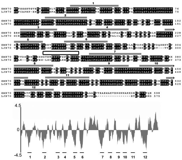
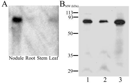
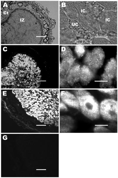
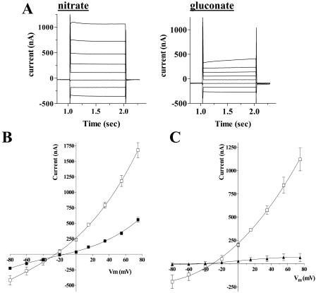
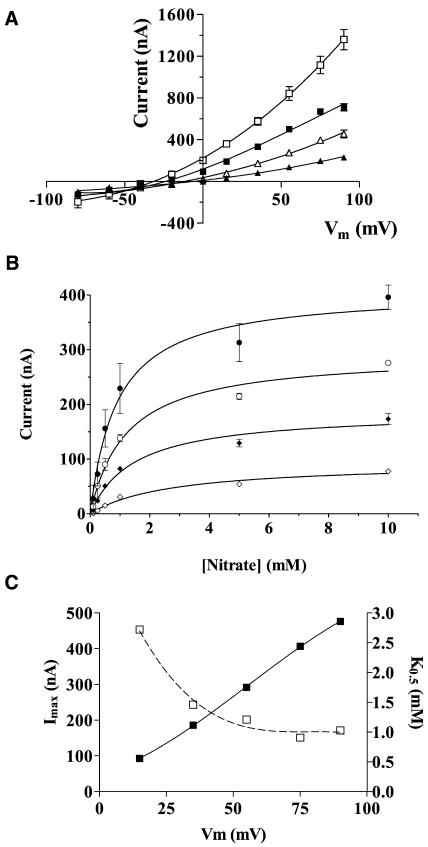
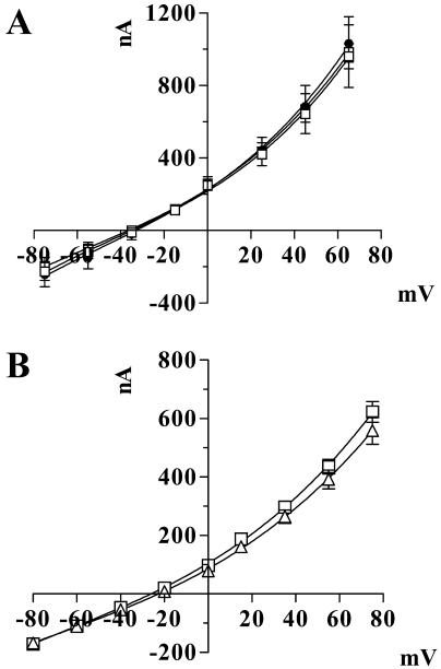
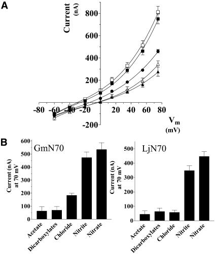
A cDNA was isolated from soybean (Glycine max) nodules that encodes a putative transporter (GmN70) of the major facilitator superfamily. GmN70 is expressed predominantly in mature nitrogen-fixing root nodules. By western-blot and immunocytochemical analyses, GmN70 was localized to the symbiosome membrane of infected root nodule cells, suggesting a transport role in symbiosis. To investigate its transport function, cRNA encoding GmN70 was expressed in Xenopus laevis oocytes, and two-electrode voltage clamp analysis was performed. Ooctyes expressing GmN70 showed outward currents that are carried by anions with a selectivity of nitrate > nitrite > > chloride. These currents showed little sensitivity to pH or the nature of the counter cation in the oocyte bath solution. One-half maximal currents were induced by nitrate concentrations between 1 to 3 mm. No apparent transport of organic anions was observed. Voltage clamp records of an ortholog of GmN70 from Lotus japonicus (LjN70; K. Szczyglowski, P. Kapranov, D. Hamburger, F.J. de Bruijn [1998] Plant Mol Biol 37: 651-661) also showed anion currents with a similar selectivity profile. Overall, these findings suggest that GmN70 and LjN70 are inorganic anion transporters of the symbiosome membrane with enhanced preference for nitrate. These transport activities may aid in regulation of ion and membrane potential homeostasis, possibly in response to external nitrate concentrations that are known to regulate the symbiosis.


Figures

Figure 1.
Figure 1.
Comparison of the deduced amino acid sequences of GmN70 and LjN70. Regions of high sequence conservation are boxed in black. Regions proposed to form transmembrane α-helices (represented by gray-shaded boxes above the sequence) were identified from a hydropathy plot (shown below the aligned sequences) by using the Kyte-Doolittle algorithm (Kyte and Doolittle, 1982) in the DNAstar software package. The sequence that was used to make a synthetic peptide antigen for site-specific antibody formation is indicated by the box between α-helices 6 and 7.
Figure 2.
Figure 2.
Northern- and western-blot analyses of GmN70. A, Total RNA samples (40 μg/lane) from nodule, root, stem, and leaf tissues of 28-d-old soybean were resolved by electrophoresis on a 1.2% (w/v) agarose-formaldahyde denaturing gel, and GmN70 mRNA was detected by northern-blot analysis as described in “Materials and Methods.” B, Purified symbiosome membranes (lanes 1 and 2) and a nodule microsomal fraction (lane 3) were resolved by SDS-PAGE on a 12.5% (w/v) polyacrylamide gel. Each lane contains 15 μg of protein. The resolved proteins were analyzed by western-blot analysis with affinity-purified anti-CD-18 antibodies as described in “Materials and Methods.”
Figure 3.
Figure 3.
Immunolocalization of GmN70 in soybean nodules. Nodule sections from 28-d-old soybeans were fixed, sectioned, and immunostained as described in “Materials and Methods.” A, Nodule section viewed by phase contrast light microscopy at low magnification with the infection (IZ) and cortical cell (Ct) zones indicated. B, A higher magnification micrograph of A, showing infected cells (IC) and uninfected companion cells (UC) of the infection zone. C and D, Nodule section probed with affinity-purified anti-GmN70 antibody. E and F, Nodule section probed with anti-nodulin-26 antibody (infected cell/symbiosome membrane control). G, Nodule section probed with preimmunization sera. The bars for A, C, E, and G are 150 μm. The bars for B, D, and F are 20 μm.
Figure 4.
Figure 4.
Nitrate-induced currents in Xenopus oocytes expressing GmN70. Oocytes were injected with GmN70 cRNA and were cultured for 4 d prior to two-electrode voltage clamp recording as described in “Materials and Methods.” A, Current records from a GmN70-injected oocyte bathed in 100 mm NaNO3 or 100 mm sodium gluconate in the presence of standard recording-bath solution. The plot represents the currents obtained from a step-wise voltage protocol in which oocytes were recorded at Vm clamped from +60 to −80 mV in 20-mV increments. Each potential was maintained for 1 s with a 0.5-s recovery period at a holding potential of −35 mV between each voltage pulse. B, Plot of steady-state currents versus clamped membrane potential (Vm) of GmN70 oocytes bathed in 100 mm Na NO3 (white squares) or 100 mm Na gluconate (black squares). Error bars show SEM of currents recorded from three separate oocytes. C, Current voltage relationship of nitrate-induced currents corrected for background currents (in the presence of gluconate) for GmN70-injected oocytes (white squares) or uninjected control ooctyes (black triangles).
Figure 5.
Figure 5.
Concentration dependence of nitrate-induced currents in Xenopus oocytes expressing GmN70. Oocytes expressing GmN70 were analyzed by voltage clamp recording under standard conditions in the presence of varying concentrations of sodium nitrate in the bath. A, I-V plots of oocytes in the presence of 100 mm (white squares), 25 mm (black squares), 10 mm (white triangles), and 1 mm (black triangles) sodium nitrate. B, Plot of steady-state currents as a function of bath nitrate concentration recorded at Vm values of +75 mV (black circles), +55 mV (white circles), +35 mV (black diamonds), and +15 mV (white diamonds). Data are the average of determinations from three oocytes with error bars showing the SEM. C, Imax (black squares) and K0.5 values (white squares) for nitrate-induced outward oocyte currents are expressed as a function of membrane potential (Vm).
Figure 6.
Figure 6.
GmN70 currents are independent of cations and pH. A, GmN70-injected oocytes were subjected to voltage clamp recording in 100 mm NO3 salts of the following cations: Na+ (white squares), K+ (black circles), and N-methyl-d-glucamine (white circles). B, Recordings of GmN70-injected oocytes in the presence of 100 mm sodium nitrate at pH 7.6 (white squares) and pH 5.0 (white triangles). The data are the average of recordings from three oocytes with error bars showing the SEM.
Figure 7.
Figure 7.
Anion selectivity of GmN70 and the L. japonicus ortholog LjN70. A, Oocytes injected with GmN70 cRNA or LjN70 cRNA were subjected to voltage clamp under standard conditions. A, GmN70-injected oocyte I-V plots in the presence of 100 mm NaNO3 (white squares), 100 mm NaNO2 (black squares), 100 mm NaCl (black circles), 66 mm sodium dicarboxylates (33 mm malate and 33 mm succinate, white triangles), and 100 mm sodium acetate (black triangles). B, Steady-state currents for GmN70 or LjN70-injected oocytes at +75 mV obtained with each of the indicated anions. The currents shown in the histograms were corrected for background currents by subtraction of basal currents obtained with gluconate substituted as the test anion. The error bars showing the SEM (n = 3 oocytes).
Figure 8.
Figure 8.
Phylogenetic analysis of GmN70-like anion transporter family with MCT-1 and OxlT-1. Shown is a phylogenetic tree of the full-length deduced amino acid sequences of the following N70-like proteins: Rice N70-like ESTs (Rice-1–7 respective accession nos.: TC235634, TC238235, NP937656, NP889385, TC217196, TC244401, TC222019); the 2 Arabidopsis N70-like proteins (accession nos. At2g39210-AAL31925, At2g28120-AAL14413); and GmN70 and LjN70. The relationship to mouse MCT-1 and O. formigenes OxlT-1 proteins (National Center for Biotechnology Information accession nos. LjN70-AAC3950, OxlT1-Q51330, MCT1-P53986) are shown. Scale bar = 0.1 amino acid substitutions/site.

Similar articles

Cited by

References

    1. Abe K, Ruan ZS, Maloney PC (1996) Cloning, sequencing, and expression in Escherichia coli of OxlT, the oxalate:formate exchange protein of Oxalobacter formigenes. J Biol Chem 271: 6789–6793 - PubMed
    1. Arrese-Igor C, Minchin FR, Gordon AJ, Nath AK (1997) Possible causes of physiological decline in soybean nitrogen fixation in the presence of nitrate. J Exp Bot 48: 905–913
    1. Barbier-Brygoo H, Vinauger M, Colcombet J, Ephritikhine G, Franchisse JM, Maurel C (2000) Anion channels in higher plants: functional characterization, molecular structure and physiological role. Biochim Biophys Acta 1465: 199–218 - PubMed
    1. Blumwald E, Fortin MG, Rea PA, Verma DPS, Poole RJ (1985) Transport of fixed nitrogen in soybean (Glycine max) root nodules. Presence of plasma membrane type H+-ATPase in the peribacteroid membrane. Plant Physiol 78: 665–672 - PMC - PubMed
    1. Brewin NJ (1991) Development of the legume root nodule. Annu Rev Cell Biol 7: 191–226 - PubMed

Publication types

MeSH terms

LinkOut - more resources