. 2005 Apr;49(4):1346-53.
doi: 10.1128/AAC.49.4.1346-1353.2005.

Development of a cell-based high-throughput specificity screen using a hepatitis C virus-bovine viral diarrhea virus dual replicon assay

Affiliations

Development of a cell-based high-throughput specificity screen using a hepatitis C virus-bovine viral diarrhea virus dual replicon assay

Donald R O'Boyle 2nd et al. Antimicrob Agents Chemother. 2005 Apr.

Abstract

The hepatitis C virus (HCV) replicon is a unique system for the development of a high-throughput screen (HTS), since the analysis of inhibitors requires the quantification of a decrease in a steady-state level of HCV RNA. HCV replicon replication is dependent on host cell factors, and any toxic effects may have a significant impact on HCV replicon replication. Therefore, determining the antiviral specificity of compounds presents a challenge for the identification of specific HCV inhibitors. Here we report the development of an HCV/bovine viral diarrhea virus (BVDV) dual replicon assay suitable for HTS to address these issues. The HCV reporter enzyme is the endogenous NS3 protease contained within the HCV genome, while the BVDV reporter enzyme is a luciferase enzyme engineered into the BVDV genome. The HTS uses a mixture of HCV and BVDV replicon cell lines placed in the same well of a 96-well plate and isolated in the same cell backgrounds (Huh-7). The format consists of three separate but compatible assays: the first quantitates the amount of cytotoxicity based upon the conversion of Alamar blue dye via cellular enzymes, while the second indirectly quantitates HCV replicon replication through measurement of the amount of NS3 protease activity present. The final assay measures the amount of luciferase activity present from the BVDV replicon cells, as an indicator of the specificity of the test compounds. This HCV/BVDV dual replicon assay provides a reliable format to determine the potency and specificity of HCV replicon inhibitors.


Figures

FIG. 1.
FIG. 1.
HCV and BVDV replicons. 5′ and 3′ indicate the cis sequence elements at the 5′ and 3′ ends of the RNA molecules necessary for viral replication. Neo is the selectable marker, EMC IRES is an IRES for internal translation of the nonstructural region of the genome, and Luc is the luciferase reporter for BVDV. A ubiquitin sequence (Ub) was placed between the luciferase and neomycin phosphotransferase genes to allow proteolytic processing. NS3 and NS4A encode the protease and activator of protease used in the cell-based FRET reporter assay for HCV, while the luciferase enzymatic activity is the reporter used for BVDV. The sites of polyprotein processing by NS3 are indicated by the arrows.
FIG. 2.
FIG. 2.
(A) Cell-based FRET assay. When HCV replicon replication is inhibited by inhibitors, the amount of HCV NS3 protease is also decreased. The amount of NS3 protease in the cell lysate then can be quantitated by the amount of the peptide substrate (AcDED[EDANS]EEAbu-y-[COO]ASK[DABCYL]-NH2) being cleaved. When the substrate is cleaved by HCV NS3 protease, it generates fluorescence. Decrease of fluorescence signal in the presence of inhibitors is due to inhibition of replicon replication. (B) Measurement of the increase in fluorescence of the HCV FRET peptide in the HCV replicon cell line and the effect of exposure to various interferon concentrations. The IFN-α concentrations (units per milliliter) used for the different wells are on the right. The assay is linear in this example over a period of 40 min.
FIG. 3.
FIG. 3.
Diagram of 96-well plate layout for HCV replicon HTS with values from Alamar blue readings and the HCV FRET assay indicated in each well. “Screen” indicates wells with test compounds; “Control” represents wells with DMSO only and is defined as 100% activity (column 11, wells A through H); “Inhibited” contains the greatest amount of a control inhibitor (100% inhibited; wells A12 and B12) and is used to determine background on each plate; “Titration” indicates the titration of interferon or a specific inhibitor and is used as a sensitivity control (wells C12 through H12). The Alamar blue reading from the random compound plate is expressed as a measure of cytotoxicity, with a low number being nontoxic. The compound in F2 shows very little toxicity, while the compound in G5 has substantial toxicity (bold outlines). The percentage of HCV protease activity in each well obtained by the calculations described in the text for the endpoint reading from cycle 21 of the FRET assay is also indicated, with low numbers being indicative of HCV replicon inhibition. The FRET numbers indicate that the compounds in wells F2 and G5 inhibited the HCV replicon ∼74% and 100%, respectively. Comparing the results of the FRET assay with the Alamar assay, it is likely that the inhibition of the HCV replicon for G5 is due to a toxic mechanism while the inhibition due to compound in F2 is not toxic in this assay, suggesting that the compound may be specific for HCV.
FIG. 4.
FIG. 4.
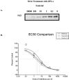
(A) Western immunoblot using an anti-NS3 protease serum for the determination of the EC50 of IFN-α. NS3 proteasespecific bands were quantified by phosphorimaging. (B) Comparison of typical values determined by FRET, qRT-PCR, or scanning of a Western blot for titration of interferon in the HCV replicon cell line. Each value represents a well of a 96-well plate at a single interferon concentration relative to a control value. Values obtained were within threefold of each other. Data at the lowest concentration of interferon tended to contain more variation.
FIG. 5.
FIG. 5.
Graphical representation of the averaged numbers from 40 separate compound plates used in the Z′ calculation. The numbers at a signal of ∼500 are the readings from the wells containing 1,000 U of IFN and are considered to have 0% FRET activity. The numbers at a signal of ∼1,500 are from wells containing buffer only and are considered to have 100% FRET activity. The Z′ measurement calculates the deviation associated with the measurements, with 2 standard deviations of the means of each measurement being acceptable (see Materials and Methods).

References

    1. Ahmed, S., J. Gopal, Jr., and J. Walsh. 1994. A new rapid and simple non-radioactive assay to monitor and determine the proliferation of lymphocytes: An alternative to H3-thymidine incorporation assay. J. Immunol. Methods 170:211-224. - PubMed
    1. Bartenschlager, R., and V. Lohmann. 2001. Novel cell culture systems for the hepatitis C virus. Antiviral Res. 52:1-17. - PubMed
    1. Behrens, S. E., C. W. Grassmann, H. J. Thiel, G. Meyers, and N. Tautz. 1998. Characterization of an autonomous subgenomic pestivirus RNA replicon. J. Virol. 72:2364-2372. - PMC - PubMed
    1. Behrens, S. E., L. Tomei, and R. De Francesco. 1996. Identification and properties of the RNA-dependent RNA polymerase of hepatitis C virus. EMBO J. 15:12-22. - PMC - PubMed
    1. Blight, K. J., A. A. Kolykhalov, and C. M. Rice. 2000. Efficient initiation of HCV RNA replication in cell culture. Science 290:1972-1974. - PubMed

Publication types

MeSH terms

LinkOut - more resources