. 2005 Apr;166(4):963-72.
doi: 10.1016/S0002-9440(10)62318-8.

Transgenic sickle mice are markedly sensitive to renal ischemia-reperfusion injury

Affiliations

Transgenic sickle mice are markedly sensitive to renal ischemia-reperfusion injury

Karl A Nath et al. Am J Pathol. 2005 Apr.

Abstract

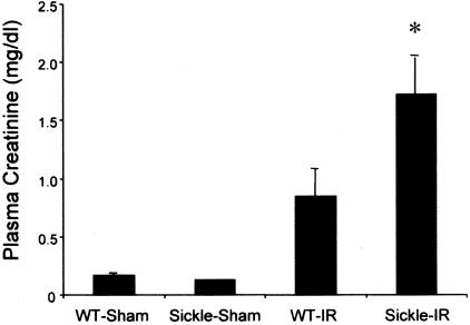
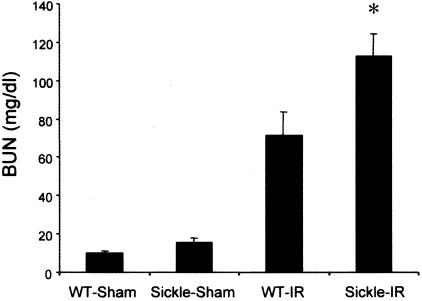
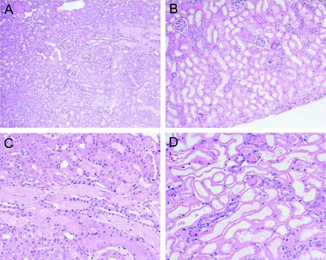
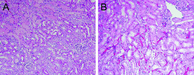
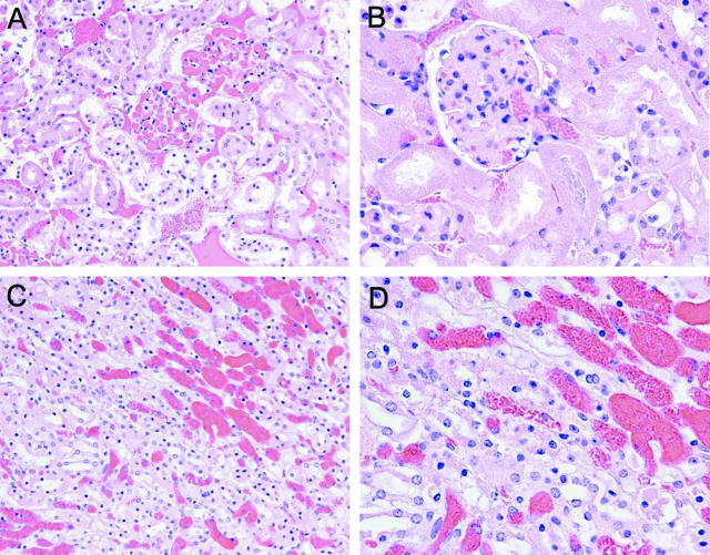
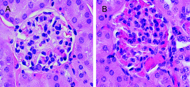
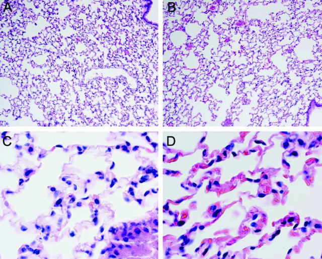
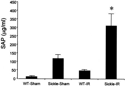
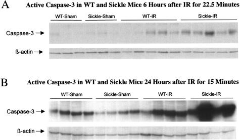
Ischemic injury is invoked as a mechanism contributing to end-organ damage and other complications of sickle cell disease (SCD). However, the intrinsic sensitivity of tissues in SCD to ischemic insults has never been addressed. We examined the effect of renal ischemia in a transgenic mouse expressing human sickle hemoglobin. Twenty-four hours after bilateral, total renal artery occlusion for 15 minutes, transgenic sickle mice exhibited worse renal function and more marked histological injury. With bilateral renal ischemia of greater duration (22.5 minutes), and after 6 hours, transgenic sickle mice exhibited massive vascular congestion, sickling of red blood cells, more marked histological injury in the kidney, and more prominent congestion in the capillary beds in the lungs and heart. Additionally, serum amyloid P-component, the murine homologue of C-reactive protein, was markedly increased in transgenic sickle mice as compared to wild-type mice. Twenty-four hours after bilateral renal ischemia for 22.5 minutes, transgenic sickle mice exhibited 28% mortality, with no mortality observed in any other group. With bilateral renal ischemia of short or long duration, renal expression of caspase-3 was most prominent in transgenic sickle mice subjected to ischemia. Thus, renal ischemia in this murine model induces more severe renal injury and extrarenal complications. We conclude that tissues in SCD exhibit heightened vascular congestion and sensitivity to ischemia and that clinically apparent or silent episodes of ischemia may contribute to the complications of SCD.


Figures

Figure 1
Figure 1
Plasma creatinine in wild-type (WT) and sickle mice subjected to bilateral renal ischemia for 15 minutes (IR) or sham-ischemia (sham), and measured 24 hours after the ischemic episode. In the sham groups n = 4 whereas n = 7 in WT-IR and n = 8 in sickle-IR. *P < 0.05 versus all other groups.
Figure 2
Figure 2
BUN in wild-type (WT) and sickle mice subjected to bilateral renal ischemia for 15 minutes (IR) or sham-ischemia (sham), and measured 24 hours after the ischemic episode. In the sham groups n = 4 whereas n = 7 in WT-IR and n = 8 in sickle-IR. *P < 0.05 versus all other groups.
Figure 3
Figure 3
Histological appearance of the kidney 24 hours after 15 minutes of bilateral renal ischemia in wild-type mice (A and C) and in sickle mice (B and D) All sections are stained by H&E. Original magnifications: ×100 (A, B); ×200 (C, D).
Figure 4
Figure 4
Histological appearance of the kidney 6 hours after 22.5 minutes of bilateral renal ischemia in wild-type mice (A) and in sickle mice (B). All sections are stained by H&E. Original magnifications, ×100.
Figure 5
Figure 5
Histological appearance of the kidney in sickle mice 6 hours after 22.5 minutes of bilateral renal ischemia. A: Cortical section demonstrating marked vascular congestion in glomerular and peritubular capillaries. B: Cortical section demonstrating marked vascular congestion and sickled RBCs in the afferent glomerular arteriole, glomerular capillaries, and peritubular capillaries; the central glomerulus shows endothelialitis and mesangiolysis, and is surrounded by necrotic proximal tubules. C: Medullary section demonstrating marked vascular congestion. D: Medullary section demonstrating marked vascular congestion and sickled RBCs as shown in the capillary in the center of the field. All sections are stained by H&E. Original magnifications: ×200 (A, C); ×400 (B, D).
Figure 6
Figure 6
Marked vascular congestion in glomerular and peritubular capillaries in the contralateral, nonclipped right kidney 6 hours after unilateral clamping of the left renal pedicle for 22.5 minutes in transgenic sickle mice (B), while vascular congestion was mild in the contralateral, nonclipped right kidney 6 hours after unilateral clamping of the left renal pedicle for 22.5 minutes in wild-type mice (A). All sections are stained by H&E. Original magnifications, ×600.
Figure 7
Figure 7
Histological appearance of the lung 6 hours after 22.5 minutes of bilateral renal ischemia in wild-type mice (A and C) and in sickle mice (B and D). All sections are stained by H&E. Original magnifications: ×100 (A, B); ×600 (C, D).
Figure 8
Figure 8
SAP in wild-type (WT) and sickle mice subjected to bilateral renal ischemia for 22.5 minutes (IR) or sham-ischemia (sham) and measured 6 hours after the ischemic episode. In the sham groups, n = 3 whereas n = 6 in WT-IR and n = 5 in sickle-IR. *P < 0.05 versus all other groups.
Figure 9
Figure 9
Expression of active caspase-3 in wild-type (WT) and sickle mice at 6 hours after bilateral renal ischemia (IR) or sham-ischemia (sham) for 22.5 minutes (A), and at 24 hours after bilateral renal ischemia (IR) or sham-ischemia (sham) for 15 minutes (B). Equivalency of loading of the Western analysis was assessed by β-actin.

References

    1. Embury SH, Hebbel RP, Steinberg MH, Mohandas N. Pathogenesis of vasoocclusion. Embury SH, Hebbel RP, Mohandas N, Steinberg MH, editors. New York: Raven,; Sickle Cell DiseaseBasic Principles and Clinical Practice. 1994:pp 311–326.
    1. Kaul DK, Fabry ME, Nagel RL. The pathophysiology of vascular obstruction in the sickle syndromes. Blood Rev. 1996;10:29–44. - PubMed
    1. Hebbel RP. Blockade of adhesion of sickle cells to endothelium by monoclonal antibodies. N Engl J Med. 2000;342:1910–1912. - PubMed
    1. Osarogiagbon UR, Choong S, Belcher JD, Vercellotti GM, Paller MS, Hebbel RP. Reperfusion injury pathophysiology in sickle transgenic mice. Blood. 2000;96:314–320. - PubMed
    1. Cheung AT, Chen PC, Larkin EC, Duong PL, Ramanujam S, Tablin F, Wun T. Microvascular abnormalities in sickle cell disease: a computer-assisted intravital microscopy study. Blood. 2002;99:3999–4005. - PubMed

Publication types