. 2005 Apr;12(4):332-9.
doi: 10.1038/nsmb916. Epub 2005 Mar 27.

Insights into hRPA32 C-terminal domain--mediated assembly of the simian virus 40 replisome

Affiliations

Insights into hRPA32 C-terminal domain--mediated assembly of the simian virus 40 replisome

Alphonse I Arunkumar et al. Nat Struct Mol Biol. 2005 Apr.

Abstract

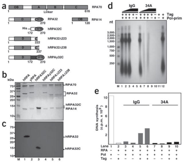
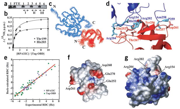
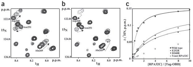
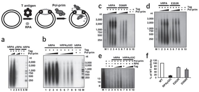
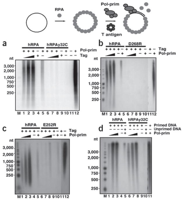
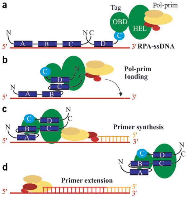
Simian virus 40 (SV40) provides a model system for the study of eukaryotic DNA replication, in which the viral protein, large T antigen (Tag), marshals human proteins to replicate the viral minichromosome. SV40 replication requires interaction of Tag with the host single-stranded DNA-binding protein, replication protein A (hRPA). The C-terminal domain of the hRPA32 subunit (RPA32C) facilitates initiation of replication, but whether it interacts with Tag is not known. Affinity chromatography and NMR revealed physical interaction between hRPA32C and the Tag origin DNA-binding domain, and a structural model of the complex was determined. Point mutations were then designed to reverse charges in the binding sites, resulting in substantially reduced binding affinity. Corresponding mutations introduced into intact hRPA impaired initiation of replication and primosome activity, implying that this interaction has a critical role in assembly and progression of the SV40 replisome.


Figures

Figure 1
Figure 1
Monoclonal antibody 34A recognizes hRPA32C and inhibits SV40 replication. (a) hRPA subunits and mutant proteins used in this study. Residue numbers are listed below each construct. OB, oligonucleotide-oligosaccharide binding folds, including the ssDNA-binding domains A– D; WH, winged helix-loop-helix domain. (b) Purified recombinant hRPA proteins were analyzed by 15% SDS-PAGE and Coomassie blue staining. M, protein markers of indicated mass. (c) Western blot assay of the proteins shown in b, probed with 34A monoclonal antibody and visualized by chemiluminescence. (d) Ab34A IgG or nonimmune mouse IgG was titrated into SV40 monopolymerase assays reconstituted with purified recombinant proteins. Products were resolved by alkaline agarose gel electrophoresis and visualized by autoradiography. Lanes 1–3 and 6–8, reactions containing 100, 300 or 500 ng of the indicated antibody. Control reactions carried out in the presence of 500 ng of antibody and in the absence of T antigen or pol-prim are indicated (−). M, DNA size marker of the indicated length in nucleotides (nt). (e) Primer synthesis and extension on M13mp18 ssDNA (25 ng) preincubated with 500 ng (lanes 3, 5, 7 and 9) or 750 ng (lanes 2, 4, 6, 8 and 10) of hRPA was tested in the presence of 250 ng pol-prim and 500 ng of either nonimmune IgG (lanes 3–6) or Ab34A (lanes 7–10). Control reactions with pol-prim alone (lane 1) and in the absence of pol-prim (lane 2) are indicated (−).
Figure 2
Figure 2
The interaction of hRPA32C with Tag-OBD. (a) Tag-OBD affinity chromatography. Lanes: I, column input; FT and E, flow-through and elution fractions from the control column; 1 and 2, flow-through and elution fractions from column I; 3 and 4, flow-through and elution fractions from column II; 5 and 6, flow-through and elution fractions from column III, respectively. The bands were quantified using Image Quant 5.0 (Molecular Dynamics). The relative amount of Tag-OBD in the elution fractions for columns I, II and III (0.25:1.00:2.75) is similar to the relative amounts of hRPA32C (0.3:0.9:3.0) used in generating the corresponding columns. All flow-through bands were saturated. (b) NMR 15N chemical shift titration curves for the binding of hRPA32C to 15N-labeled Tag-OBD. The changes in amide nitrogen chemical shifts of Thr199 and His203 in Tag are plotted against the ratio of Tag-OBD to hRPA32C. The line through each curve represents a best fit to the standard single-site binding equation. (c) Ensemble of 20 lowest-energy conformers of the complex of Tag (blue) and hRPA32C (red). (d) Side chains in the binding interface of the representative Tag-OBD–RPA32C structure. Tag-OBD is blue and hRPA32C is red. The side chains are not as well defined as the backbone, so conclusions should not be drawn regarding specific intermolecular interactions apparent in this figure showing only a single conformer. (e) Correlation between the experimentally measured 1DNH residual dipolar couplings (RDC) versus the values back-calculated from the representative structure. (f,g) Electrostatic surfaces of the two molecules in the Tag-OBD–RPA32C complex. Red and blue, negative and positive charge, respectively. Key residues in the binding interface are labeled.
Figure 3
Figure 3
Effects of DNA binding and mutations on the interaction between hRPA32C and Tag-OBD. (a,b) Comparison of the binding of Tag-OBD to hRPA32C in the absence (a) and presence (b) of origin DNA. Unlabeled Tag-OBD was titrated into a 100 mM solution of 15N-enriched hRPA32C and a series of 15N,1H HSQC NMR spectra were acquired. An overlay of a small region from these spectra is shown in a. A stoichiometric amount of origin DNA duplex was then titrated into the solution and an additional spectrum was acquired (b). Arrows facilitate following the change in the location of the NMR signal. (c) NMR 1H chemical shift titration curves for the binding of wild-type, E252R, E268R and yeast hRPA32C to 15N-labeled Tag-OBD. The changes in amide proton chemical shifts of Thr199 are plotted against the ratio of Tag-OBD to hRPA32C. The line through each curve represents a best fit to the standard single-site binding equation.
Figure 4
Figure 4
Mutations in hRPA32C that weaken interaction with Tag are defective in initiation of SV40 DNA replication. (a–d) Initiation of replication was tested in monopolymerase reactions containing 200 ng of the indicated hRPA and 300 or 400 (a), or 100–400 ng (bd), of pol-prim as indicated. Control reactions contained hRPA but lacked either pol-prim or Tag as indicated (−). The products were resolved by alkaline agarose gel electrophoresis and visualized by autoradiography. DNA size markers are indicated (M). (e) SV40 monopolymerase reactions containing radiolabeled CTP were carried out in the presence of 200 ng of the indicated hRPAs, 250 ng of pol-prim, and 250–750 ng of Tag as indicated. Control reactions lacking Tag, hRPA or pol-prim are indicated (−). Radiolabeled RNA products were resolved by electrophoresis on a polyacrylamide gel containing 20% urea and visualized by autoradiography. M, radiolabeled oligonucleotide size marker dT (4–22). Arrowhead indicates RNA primers of 8–10 nt. (f) Primer synthesis in the monopolymerase reaction was quantified and expressed as a percentage of wild-type activity. At least two reactions were used for quantification of each mutant. Brackets represent standard error.
Figure 5
Figure 5
hRPA32C is needed for primosome activity, but not for primer extension. (a–c) Primer synthesis and extension was assayed on 100 ng M13 ssDNA precoated with 600 ng of hRPA or mutant hRPA as indicated. Reactions contained 250 ng of pol-prim and 250–750 ng of Tag as indicated. Control reactions lacked hRPA, pol-prim or Tag as indicated (−). Radiolabeled DNA products were resolved by alkaline agarose gel electrophoresis and visualized by autoradiography. M, DNA size markers as indicated. (d) Singly primed ssDNA (100 ng) precoated with 1,000 ng of the indicated hRPA was incubated with purified pol-prim (100, 150 and 250 ng) as indicated (+). Negative control reactions were done without pol-prim (lanes 4 and 9) or with unprimed template (lanes 5 and 10). Primer extension in the absence of hRPA is shown in lane 11.
Figure 6
Figure 6
Model for SV40 primosome activity on hRPA-coated ssDNA. (a) hRPA (blue) is schematically depicted in the high-affinity 28–30 nt binding mode with all four ssDNA binding domains (A–D) bound to ssDNA. hRPA14 is omitted for simplicity. The helicase domain (HEL) of a Tag hexamer (green) can associate with a pol-prim heterotetramer,,. Antibodies against Tag that specifically inhibit either hRPA binding to Tag-OBD or pol-prim binding to the helicase domain prevent primer synthesis. (b) We suggest that primosome assembly begins when Tag-OBD associates first with hRPA32C and then with hRPA70AB, transiently creating a short stretch of unbound ssDNA. (c) In concert with this hRPA remodeling, pol-prim associated with the Tag hexamer would be poised to access the free ssDNA and begin primer synthesis. (d) Primer extension by pol-prim is likely coupled with hRPA and Tag dissociation, and followed by the RFC/PCNA-mediated switch to DNA polymerase δ (not shown).

References

    1. Fanning E, Knippers R. Structure and function of simian virus 40 large tumor antigen. Annu Rev Biochem. 1992;61:55–85. - PubMed
    1. Bullock PA. The initiation of simian virus 40 DNA replication in vitro. Crit Rev Biochem Mol Biol. 1997;32:503–568. - PubMed
    1. Simmons DT. SV40 large T antigen functions in DNA replication and transformation. Adv Virus Res. 2000;55:75–134. - PubMed
    1. Stenlund A. Initiation of DNA replication: lessons from viral initiator proteins. Nat Rev Mol Cell Biol. 2003;4:777–785. - PubMed
    1. Stauffer ME, Chazin WJ. Structural mechanisms of DNA replication, repair, and recombination. J Biol Chem. 2004;279:30915–30918. - PubMed

Publication types

MeSH terms

Associated data