. 2005 Apr;79(8):4764-73.
doi: 10.1128/JVI.79.8.4764-4773.2005.

Expression of matrix metalloproteinases and their tissue inhibitor during viral encephalitis

Affiliations

Expression of matrix metalloproteinases and their tissue inhibitor during viral encephalitis

Jiehao Zhou et al. J Virol. 2005 Apr.

Abstract

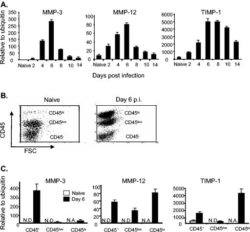
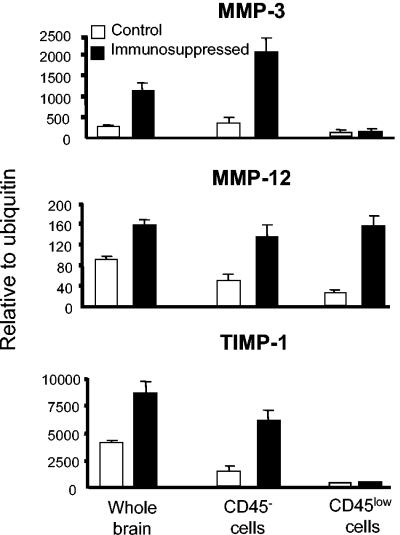
Matrix metalloproteinases (MMPs) participate in remodeling the extracellular matrix and facilitate entry of inflammatory cells into tissues. Infection of the murine central nervous system (CNS) with a neurotropic coronavirus induces encephalitis associated with increased levels of mRNA encoding MMP-3 and MMP-12. Whereas virus-induced MMP-3 expression was restricted to CNS resident astrocytes, MMP-12 mRNA was expressed by both inflammatory cells and CNS resident cells. Immunosuppression increased both MMP-3 and MMP-12 mRNA levels in CNS resident cells, suggesting that the presence of virus rather than inflammation induced protease up-regulation. MMP activity is partially regulated by a small family of genes encoding tissue inhibitors of matrix metalloproteinases (TIMPs); among the TIMPs, only TIMP-1 mRNA expression increased in the CNS following coronavirus infection. During inflammation TIMP-1 mRNA was most prominently expressed by infiltrating cells. By contrast, in the immunosuppressed host TIMP-1 mRNA was expressed by CNS resident cells. Analysis of cytokine and chemokine mRNA induction within the infected CNS of healthy and immunocompromised mice suggested a possible correlation between increased viral replication and increased levels of beta interferon, MMP-3, MMP-12, and TIMP-1 mRNA. CD4+ T cells which localize to the perivascular and subarachnoid spaces were identified as the primary source of TIMP-1 protein. By contrast, protein expression was undetectable in astrocytes or CD8+ T cells, the primary antiviral effectors that localize to the CNS parenchyma in response to infection. These data suggest that in contrast to the results seen with MMPs, inhibition of protease activity via TIMP-1 expression correlates with the differential tissue distribution of T-cell subsets during acute coronavirus-induced encephalitis.


Figures

FIG. 1.
FIG. 1.
Selective up-regulation of MMP-3, MMP-12, and TIMP-1 mRNA during viral encephalitis. (A) MMP-3, MMP-12, and TIMP-1 mRNA expression in brains of naïve and infected mice determined by semiquantitative real-time PCR. Data representing the average ± SD for three to five mice per group are presented in units relative to ubiquitin mRNA expression levels. (B) Flow cytometric analysis of CD45-expressing brain cells isolated from naïve or infected mice at day 6 p.i. (C) Comparison of MMP-3, MMP-12, and TIMP-1 mRNA expression in the CD45 CNS resident cells, CD45low microglia, and CD45hi bone-marrow-derived cells from infected mice at day 6 p.i. Data represent the average results of three separate experiments. Error bars represent SD. N.D., not detectable. N.A., not applicable.
FIG. 2.
FIG. 2.
Absence of inflammatory cells and increased infectious virus following immunosuppression. (A) Flow cytometric analysis of CNS-derived cells prepared from JHMV-infected immunosuppressed mice and wild-type controls at day 6 p.i. (A) Note the absence of CD45hi bone-marrow-derived inflammatory cells and retention of CD45low microglia. (B) Increased infectious virus levels in the CNS of immunosuppressed versus untreated infected mice. Data represent the average of duplicate determinations for a minimum of three mice in each group.
FIG. 3.
FIG. 3.
Enhanced MMP-3, MMP-12, and TIMP-1 mRNA expression in the CNS of immunosuppressed hosts following infection. The results of a comparison of the levels of MMP-3, MMP-12, and TIMP-1 mRNA expression in unfractionated brain, CD45 CNS resident cells, and CD45low microglia derived from untreated infected and immunosuppressed infected mice at day 6 p.i. are shown. Data represent average results of two separate experiments. Error bars represent ranges of experimental results.
FIG. 4.
FIG. 4.
Expression of cytokines and chemokines in the brains of infected untreated and immunosuppressed infected mice. Relative levels of mRNA expression of cytokine (A) and chemokine (B) mRNA in brains of untreated control and immunosuppressed infected mice at day 6 p.i. are shown. Data represent the average results obtained for three to five mice per group. Error bars represent SD. *, P < 0.05.
FIG. 5.
FIG. 5.
Astrocyte expression of MMP-3 during virus-induced encephalitis. (A) Expression of MMP-3 mRNA in CNS resident and inflammatory cells isolated by FACS at day 6 p.i. Samples were pooled from four to seven mice/experiment. mRNA levels determined by real-time PCR are presented relative to ubiquitin mRNA expression levels. Data represent the average results of three experiments. (B) MMP-3 protein expression in the infected CNS. Arrows indicate cells giving positive results. (C) MMP-3 colocalizes with GFAP by confocal microscopy. MMP-3-expressing cells were detected with a tetramethyl rhodamine isothiocyanate conjugate (red). GFAP positive cells were detected with an FITC conjugate (green). Cells coexpressing MMP-3 and GFAP are yellow.
FIG. 6.
FIG. 6.
MMP-12 mRNA expression in CNS-derived cells. CNS resident and inflammatory cells were isolated by FACS from pools of four to seven infected mice/experiment at day 6 p.i. Data represent the average results of three experiments. MMP-12 mRNA levels were determined by real-time PCR and are presented relative to ubiquitin mRNA expression levels.
FIG. 7.
FIG. 7.
TIMP-1 expression by CD4+ T cells. (A) CNS resident and inflammatory cells isolated by FACS were pooled from four to seven infected mice/experiment at day 6 p.i. Data represent the average results of three experiments. TIMP-1 mRNA levels were determined by real-time PCR are presented relative to ubiquitin mRNA expression levels. (B) Distribution of TIMP-1, CD4+ T, and CD8+ T cells in brains of infected mice. (C) Colocalization of TIMP-1 and CD4. TIMP-1-expressing cells were detected with a Cy3 conjugate (red). CD4+ T cells were detected with an FITC conjugate (green). Cells coexpressing TIMP-1 and CD4 are yellow.
FIG. 8.
FIG. 8.
MMP and TIMP expression in naïve versus activated CD4+ T cells. CD4+ T cells were purified by positive selection from BALB/c mice and activated with PMA and ionomycin for 12 h. MMP and TIMP mRNA levels in untreated and activated CD4+ T cells were determined by real-time PCR and are presented relative to ubiquitin mRNA expression levels.

References

    1. Alter, A., M. Duddy, S. Herbert, K. Biernacki, A. Prtat, J. Antel, V. W. Yong, R. K. Nuttall, C. J. Pennington, D. R. Edwards, and A. Bar-Or. 2003. Determinants of human B cell migration across brain endothelial cells. J. Immunol. 170:4497-4505. - PubMed
    1. Bart, J., H. J. Groen, N. K. Hendrikse, W.T. van der Graaf, W. Vaalburg, and E. G. de Vries. 2000. The blood-brain barrier and oncology: new insights into function and modulation. Cancer Treat. Rev. 26:449-462. - PubMed
    1. Bergmann, C., B. Parra, D. R. Hinton, C. Ramakrishna. M. Morrison, and S. A. Stohlman. 2003. Perforin mediated effector function within the CNS requires IFN-g mediated MHC upregulation. J. Immunol. 170:3204-3213. - PubMed
    1. Brosnan, C. F., W. Cammer, W. T. Norton, and R. Bloom. 1980. Proteinase inhibitors suppress the development of experimental allergic encephalomyelitis. Nature 285:235-237. - PubMed
    1. Bugnu, M., B. Witek, J. Bereta, M. Bereta, D. R. Edwards, and T. Kordula. 1999. Reprogramming of TIMP-1 and TIMP-3 expression profiles in brain microvascular endothelial cells and astrocytes in response to proinflammatory cytokines. FEBS Lett. 448:9-14. - PubMed

Publication types