. 2005 Apr;79(8):4896-907.
doi: 10.1128/JVI.79.8.4896-4907.2005.

CD4+ T-cell responses to Epstein-Barr virus (EBV) latent-cycle antigens and the recognition of EBV-transformed lymphoblastoid cell lines

Affiliations
Free PMC article

CD4+ T-cell responses to Epstein-Barr virus (EBV) latent-cycle antigens and the recognition of EBV-transformed lymphoblastoid cell lines

H M Long et al. J Virol. 2005 Apr.
Free PMC article

Abstract

There is considerable interest in the potential of Epstein-Barr virus (EBV) latent antigen-specific CD4+ T cells to act as direct effectors controlling EBV-induced B lymphoproliferations. Such activity would require direct CD4+ T-cell recognition of latently infected cells through epitopes derived from endogenously expressed viral proteins and presented on the target cell surface in association with HLA class II molecules. It is therefore important to know how often these conditions are met. Here we provide CD4+ epitope maps for four EBV nuclear antigens, EBNA1, -2, -3A, and -3C, and establish CD4+ T-cell clones against 12 representative epitopes. For each epitope we identify the relevant HLA class II restricting allele and determine the efficiency with which epitope-specific effectors recognize the autologous EBV-transformed B-lymphoblastoid cell line (LCL). The level of recognition measured by gamma interferon release was consistent among clones to the same epitope but varied between epitopes, with values ranging from 0 to 35% of the maximum seen against the epitope peptide-loaded LCL. These epitope-specific differences, also apparent in short-term cytotoxicity and longer-term outgrowth assays on LCL targets, did not relate to the identity of the source antigen and could not be explained by the different functional avidities of the CD4+ clones; rather, they appeared to reflect different levels of epitope display at the LCL surface. Thus, while CD4+ T-cell responses are detectable against many epitopes in EBV latent proteins, only a minority of these responses are likely to have therapeutic potential as effectors directly recognizing latently infected target cells.


Figures

FIG. 1.
FIG. 1.
Mapping of CD4+ epitopes within the primary sequences of EBNA1, -2, -3A, and -3C. The position of each epitope is identified by a number that refers to the coordinate of the first amino acid in the protein as encoded by EBV strain B95.8. The bar above each epitope position indicates the percentage of all EBV-seropositive donors tested who responded to the relevant epitope peptide in ELISPOT assays. Epitopes against which CD4+ T-cell clones were later derived are also identified by a three-letter code corresponding to the first three amino acids of the epitope sequence and by an asterisk above the bar. For each protein, the number of unique amino acids (aa) is shown; this excludes the 233-amino-acid glycine-alanine repeat domain (residues 93 to 325) for EBNA1 and the 42-amino-acid polyproline repeat domain (residues 59 to 100) for EBNA2.
FIG. 2.
FIG. 2.
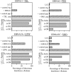
Antigen (Ag) specificity of CD4+ T-cell clones raised against the TSL (EBNA1 515), PRS (EBNA2 276), GPW (EBNA3A 780), and SDD (EBNA3C 386) epitopes. (Top panels) Clones (500 T cells per well) were stimulated overnight with autologous LCLs that were either unmanipulated, prepulsed for 1 h with 5 μM epitope peptide (pep; or an equivalent concentration of control peptide), or preexposed in serum-free medium for 2 h to specific EBV antigen preparations (or to a control antigen) and then washed before the assay. Responses are expressed as percentages of the maximum IFN-γ induced by peptide-loaded target cells in each case. (Bottom panels) The results shown in the bottom panels are from a separate experiment in which autologous LCLs were preexposed for 1 h to epitope peptide at a concentration mediating half-maximal responses, washed, and then incubated for a further hour in the presence of MAb to HLA-DP, HLA-DQ, or HLA-DR or in medium as a control (No MAb) before the addition of 500 T cells to the cell suspension. Results are expressed as described above. Note that assays conducted with the PRS-specific clones were carried out with LCLs transformed with the type 2 Ag876 EBV strain, in which the PRS epitope sequence is mutated (10), thereby reducing background LCL recognition to zero.
FIG. 3.
FIG. 3.
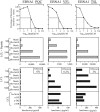
Functional analysis of CD4+ T-cell clones against the PQC (EBNA1 529), VFL (EBNA1 564), and TSL (EBNA1 515) epitopes. (Top panels) Clones (100 T cells per well) were stimulated overnight with autologous LCLs (5 × 10⁴ per well) either unmanipulated (neg) or loaded with epitope peptide at 10−5 to 10−10 M concentrations. Responses were assayed by IFN-γ release and expressed as a percentage of the maximum peptide-induced response. (Middle panels) Clones (500 to 5,000 T cells per well) were stimulated as described above with the autologous LCLs (match) or with HLA class II-mismatched LCLs (5 × 10⁴ per well), both previously exposed to 5 μM epitope peptide and then washed before the assay. Responses are expressed as IFN-γ release in picograms per milliliter. (Bottom panels, upper section) Clones were tested at the same time as above, on the same autologous and HLA class II-mismatched LCLs but with no exogenous peptide treatment. Responses are expressed as IFN-γ release in picograms per milliliter. The efficiency with which each clone is able to recognize unmanipulated autologous LCL targets is expressed as a percentage of the maximal response seen on the same targets loaded with peptide (box at upper right of each graph). (Bottom panels, lower section) The results shown in the lower section of the bottom panels are from a separate experiment in which the responses of 500 T cells to the autologous LCL (non-peptide loaded) were assayed either alone (no MAb) or in the presence of MAbs to HLA-DP, HLA-DQ, or HLA-DR as described above.
FIG. 4.
FIG. 4.
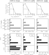
Functional analysis of CD4+ T-cell clones against the PAQ (EBNA2, 301), GPW (EBNA3A, 780), and SDD (EBNA3C, 386) epitopes. The experimental design and expression of results are the same as in Fig. 3, except that the range of T-cell numbers used in LCL stimulation experiments extended from 100 to 2,500 per well.
FIG. 5.
FIG. 5.
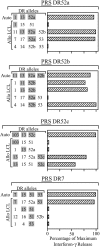
Analysis of HLA class II allele restriction of CD4+ T-cell clones specific for the PRS (EBNA2, 276) epitope. Clones were established from four donors whose HLA-DR types are in each case identified (Auto). These clones were then tested as described in the legend to Fig. 3 (middle panels) against peptide-loaded cells of the autologous LCL and of allogeneic LCLs of known HLA-DR type (Allo LCL). Alleles matched with the autologous cells are identified by shading. Results are expressed as a percentage of the maximum IFN-γ release observed in the assay.
FIG. 6.
FIG. 6.
Functional analysis of CD4+ T-cell clones specific for the PRS (EBNA2, 276) epitope derived from different EBV-seropositive donors and restricted through HLA-DR52a, -DR52b, -DR52c, and -DR7, respectively. The experimental design and expression of results are the same as in Fig. 4. Note that peptide titration assays involving the DR52b-restricted PRS clones were conducted with the Ag876 virus-transformed LCL as in Fig. 2.
FIG. 7.
FIG. 7.
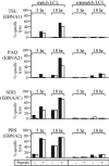
Killing of LCL targets by CD4+ T-cell clones against the TSL (EBNA1 515), PAQ (EBNA2 301), SDD (EBNA3C 386), and PRS (EBNA2 276) epitopes. Five- and 18-h chromium release assays were conducted with HLA class II-matched and mismatched LCL targets either unmanipulated or previously exposed to 5 μM epitope peptide and then washed before the assay. Results are expressed as percent specific chromium release from target cells at effector/target ratios of 5:1 (▪) and 2.5:1 (□).
FIG. 8.
FIG. 8.
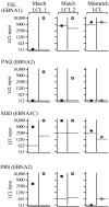
Inhibition of LCL outgrowth by the epitope-specific CD4+ T-cell clones used in Fig. 7. For each clone, two HLA class II-matched LCLs and one mismatched LCL were seeded at doubling dilutions of 104 to 300 cells per well either alone or with the addition of 104 CD4+ T cells. The LCLs were either unmanipulated or previously exposed to 5 μM epitope peptide and then washed before seeding. Results are expressed as the minimum LCL seeding required for successful outgrowth in each case. For each clone, the results from LCL-T-cell cocultures are shown for the unmanipulated LCL (▪) and in the adjacent column for the peptide-loaded LCL (░⃞). These values are in each case compared with the corresponding results for outgrowth of the unmanipulated LCL or of the peptide-loaded LCL cultured in the absence of T cells (dotted lines).

References

    1. Babcock, G. J., L. L. Decker, M. Volk, and D. A. Thorley-Lawson. 1998. EBV persistence in memory B cells in vivo. Immunity 9:395-404. - PubMed
    1. Chen, M., M. Shirai, Z. Liu, T. Arichi, H. Takahashi, and M. Nishioka. 1998. Efficient class II major histocompatibility complex presentation of endogenously synthesized hepatitis C virus core protein by Epstein-Barr virus-transformed B-lymphoblastoid cell lines to CD4+ T cells. J. Virol. 72:8301-8308. - PMC - PubMed
    1. Crotzer, V. L., R. E. Christian, J. M. Brooks, J. Shabanowitz, R. E. Settlage, J. A. Marto, F. M. White, A. B. Rickinson, D. F. Hunt, and V. H. Engelhard. 2000. Immunodominance among EBV-derived epitopes restricted by HLA-B27 does not correlate with epitope abundance in EBV-transformed B-lymphoblastoid cell lines. J. Immunol. 164:6120-6129. - PubMed
    1. Greenspan, J. S., D. Greenspan, E.T. Lennette, D. I. Abrams, M. A. Conant, V. Petersen, and U. K. Freese. 1985. Replication of Epstein-Barr virus within the epithelial cells of oral “hairy” leukoplakia, an AIDS-associated lesion. N. Engl. J. Med. 313:1564-1571. - PubMed
    1. Gudgeon, N. H., G. S. Taylor, H. M. Long, T. A. Haigh, and A. B. Rickinson. Regression of Epstein-Barr virus-induced B-cell transformation in vitro involves virus-specific CD8+T cells as the principal effectors and a novel CD4+ T-cell reactivity J. Virol., in press. - PMC - PubMed

Publication types