CD4-independent entry and replication of simian immunodeficiency virus in primary rhesus macaque astrocytes are regulated by the transmembrane protein
- PMID: 15795280
- PMCID: PMC1069519
- DOI: 10.1128/JVI.79.8.4944-4951.2005
CD4-independent entry and replication of simian immunodeficiency virus in primary rhesus macaque astrocytes are regulated by the transmembrane protein
Abstract
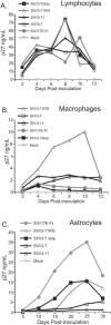
Previous studies have demonstrated that the genetic determinants of simian immunodeficiency virus (SIV) neurovirulence map to the env and nef genes. Recent studies from our laboratory demonstrated that SIV replication in primary rhesus macaque astrocyte cultures is dependent upon the nef gene. Here, we demonstrate that macrophage tropism is not sufficient for replication in astrocytes and that specific amino acids in the transmembrane (TM) portion of Env are also important for optimal SIV replication in astrocytes. Specifically, a Gly at amino acid position 751 and truncation of the cytoplasmic tail of TM are required for efficient replication in these cells. Studies using soluble CD4 demonstrated that these changes within the TM protein regulate CD4-independent, CCR5-dependent entry of virus into astrocytes. In addition, we observed that two distinct CD4-independent, neuroinvasive strains of SIV/DeltaB670 also replicated efficiently in astrocytes, further supporting the role of CD4 independence as an important determinant of SIV infection of astrocytes in vitro and in vivo.
Figures


Similar articles
-
Expression of simian immunodeficiency virus (SIV) nef in astrocytes during acute and terminal infection and requirement of nef for optimal replication of neurovirulent SIV in vitro.J Virol. 2003 Jun;77(12):6855-66. doi: 10.1128/jvi.77.12.6855-6866.2003. J Virol. 2003. PMID: 12768005 Free PMC article.
-
A single amino acid change and truncated TM are sufficient for simian immunodeficiency virus to enter cells using CCR5 in a CD4-independent pathway.Virology. 2005 Oct 10;341(1):12-23. doi: 10.1016/j.virol.2005.07.001. Virology. 2005. PMID: 16061266 Free PMC article.
-
Substitutions in Nef That Uncouple Tetherin and SERINC5 Antagonism Impair Simian Immunodeficiency Virus Replication in Primary Rhesus Macaque Lymphocytes.J Virol. 2022 Jun 8;96(11):e0017622. doi: 10.1128/jvi.00176-22. Epub 2022 May 10. J Virol. 2022. PMID: 35536019 Free PMC article.
-
Derivation and Characterization of a CD4-Independent, Non-CD4-Tropic Simian Immunodeficiency Virus.J Virol. 2016 Apr 29;90(10):4966-4980. doi: 10.1128/JVI.02851-15. Print 2016 May 15. J Virol. 2016. PMID: 26937037 Free PMC article.
-
Immunodeficiency viruses. Not enough sans Nef.Curr Biol. 1994 Jul 1;4(7):618-20. doi: 10.1016/s0960-9822(00)00135-4. Curr Biol. 1994. PMID: 7953537 Review.
Cited by
-
HIV infection of non-classical cells in the brain.Retrovirology. 2023 Jan 13;20(1):1. doi: 10.1186/s12977-023-00616-9. Retrovirology. 2023. PMID: 36639783 Free PMC article. Review.
-
SAMHD1 transcript upregulation during SIV infection of the central nervous system does not associate with reduced viral load.Sci Rep. 2016 Mar 3;6:22629. doi: 10.1038/srep22629. Sci Rep. 2016. PMID: 26936683 Free PMC article.
-
Induction of antibody-mediated neutralization in SIVmac239 by a naturally acquired V3 mutation.Virology. 2010 Apr 25;400(1):86-92. doi: 10.1016/j.virol.2010.01.014. Epub 2010 Feb 11. Virology. 2010. PMID: 20153009 Free PMC article.
-
Laser capture microdissection assessment of virus compartmentalization in the central nervous systems of macaques infected with neurovirulent simian immunodeficiency virus.J Virol. 2013 Aug;87(16):8896-908. doi: 10.1128/JVI.00874-13. Epub 2013 May 29. J Virol. 2013. PMID: 23720733 Free PMC article.
-
Canonical type I IFN signaling in simian immunodeficiency virus-infected macrophages is disrupted by astrocyte-secreted CCL2.J Immunol. 2012 Apr 15;188(8):3876-85. doi: 10.4049/jimmunol.1103024. Epub 2012 Mar 9. J Immunol. 2012. PMID: 22407919 Free PMC article.
References
-
- Anderson, M. G., D. Hauer, D. P. Sharma, S. V. Joag, O. Narayan, M. C. Zink, and J. E. Clements. 1993. Analysis of envelope changes acquired by SIVmac239 during neuroadaption in rhesus macaques. Virology 195:616-626. - PubMed
-
- Babas, T., E. Vieler, D. A. Hauer, R. J. Adams, P. M. Tarwater, K. Fox, J. E. Clements, and M. C. Zink. 2001. Pathogenesis of SIV pneumonia: selective replication of viral genotypes in the lung. Virology 287:371-381. - PubMed
-
- Barber, S. A., Herbst, D. S., Bullock, B. T., Gama, L., and J. E. Clements. 2004. Innate immune response and control of acute simian immunodeficiency virus replication in the central nervous system. J. Neurovirol. 10:15-20. - PubMed
Publication types
MeSH terms
Substances
Grants and funding
LinkOut - more resources
Full Text Sources
Research Materials

