

Plateau levels of viremia correlate with the degree of CD4+-T-cell loss in simian immunodeficiency virus SIVagm-infected pigtailed macaques: variable pathogenicity of natural SIVagm isolates
- PMID: 15795299
- PMCID: PMC1069563
- DOI: 10.1128/JVI.79.8.5153-5162.2005
Plateau levels of viremia correlate with the degree of CD4+-T-cell loss in simian immunodeficiency virus SIVagm-infected pigtailed macaques: variable pathogenicity of natural SIVagm isolates
Abstract
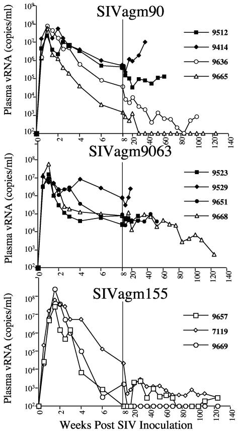
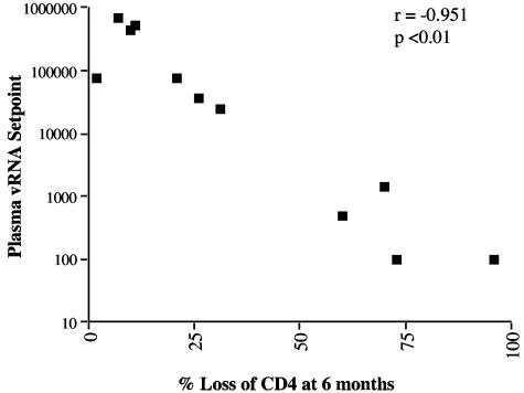
Simian immunodeficiency virus from African green monkeys (SIVagm) results in asymptomatic infection in its natural host species. The virus is not inherently apathogenic, since infection of pigtailed (PT) macaques (Macaca nemestrina) with one isolate of SIVagm results in an immunodeficiency syndrome characterized by progressive CD4+-T-cell depletion and opportunistic infections. This virus was passaged once in a PT macaque and, thus, may not be entirely reflective of the virulence of the parental strain. The goal of the present study was to assess the pathogenicity of the PT-passaged isolate (SIVagm9063) and two primary SIVagm isolates in PT macaques, including the parental strain of the PT-passaged variant. Infection of macaques with any of the three isolates resulted in high levels of primary plasma viremia by 1 week after inoculation. Viremia was quickly controlled following infection with SIVagm155; these animals have maintained CD4+-T-cell subsets and remain healthy. The plateau levels among SIVagm90- and SIVagm9063-inoculated macaques varied widely from 100 to 1 million copies/ml of plasma. Three of four animals from each of these groups progressed to AIDS. Setpoint viremia and the degree of CD4+-T-cell loss at 6 months postinfection were not significantly different between macaques inoculated with SIVagm90 and SIVagm9063. However these parameters were significantly different in SIVagm155-inoculated macaques (P values of <0.01). Considering all the macaques, the degree of CD4+-T-cell loss by 6 months postinfection correlated with the plateau levels of viremia. Thus, similar to SIVsm/mac infection of macaques and human AIDS, viral load is an excellent prognostic indicator of disease course. The inherent pathogenicity of natural SIVagm isolates varies, but such natural isolates are capable of inducing AIDS in macaques without prior macaque passage.
Figures


References
-
- Apetrei, C., D. L. Robertson, and P. A. Marx. 2004. The history of SIVS and AIDS: epidemiology, phylogeny and biology of isolates from naturally SIV infected non-human primates (NHP) in Africa. Front. Biosci. 9:225-254. - PubMed
-
- Baier, M., A. Werner, K. Cichutek, C. Garber, C. Muller, G. Kraus, F. J. Ferdinand, S. Hartung, T. S. Papas, and R. Kurth. 1989. Molecularly cloned simian immunodeficiency virus SIVagm3 is highly divergent from other SIVagm isolates and is biologically active in vitro and in vivo. J. Virol. 63:5119-5123. - PMC - PubMed
-
- Bailes, E., F. Gao, F. Bibollet-Ruche, V. Courgnaud, M. Peeters, P. A. Marx, B. H. Hahn, and P. M. Sharp. 2003. Hybrid origin of SIV in chimpanzees. Science 300:1713. - PubMed
-
- Baskin, G. B., L. N. Martin, M. Murphey-Corb, F. S. Hu, D. Kuebler, and B. Davison. 1995. Distribution of SIV in lymph nodes of serially sacrificed rhesus monkeys. AIDS Res. Hum. Retrovir. 11:273-285. - PubMed
MeSH terms
Substances
LinkOut - more resources
Full Text Sources
Research Materials
