. 2005 Apr;79(8):5174-84.
doi: 10.1128/JVI.79.8.5174-5184.2005.

Comparison of inflammatory and acute-phase responses in the brain and peripheral organs of the ME7 model of prion disease

Affiliations

Comparison of inflammatory and acute-phase responses in the brain and peripheral organs of the ME7 model of prion disease

Colm Cunningham et al. J Virol. 2005 Apr.

Abstract

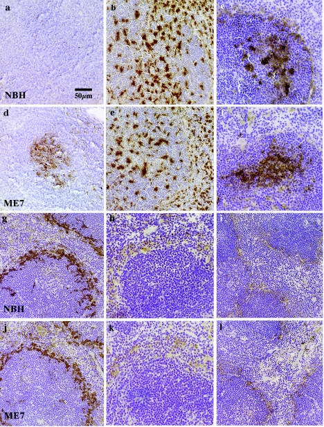
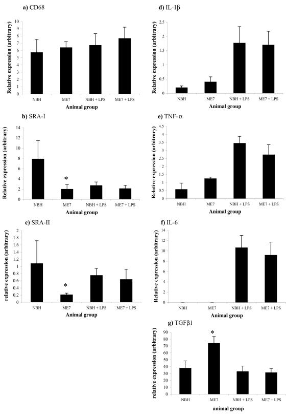
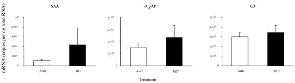
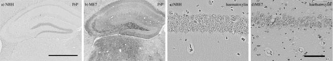
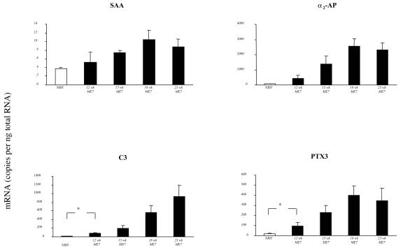
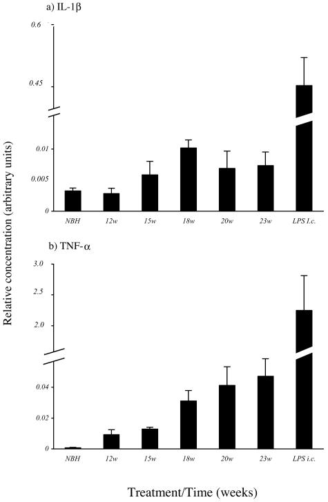
Chronic neurodegenerative diseases such as prion disease and Alzheimer's disease (AD) are reported to be associated with microglial activation and increased brain and serum cytokines and acute-phase proteins (APPs). Unlike AD, prion disease is also associated with a peripheral component in that the presumed causative agent, PrPSc, also accumulates in the spleen and other lymphoreticular organs. It is unclear whether the reported systemic acute-phase response represents a systemic inflammatory response to prion disease or merely reflects central nervous system (CNS) inflammation. For this study, we investigated whether intracerebrally initiated prion disease (ME7 model) provokes splenic, hepatic, or brain inflammatory and acute-phase responses. We detected no significant elevation of proinflammatory cytokines or activation of macrophages in the spleens of these animals, despite clear PrPSc deposition. Similarly, at 19 weeks we detected no significant elevation of transcripts for the APPs serum amyloid A, complement C3, pentraxin 3, and alpha2-antiplasmin in the liver, despite CNS neurodegeneration and splenic PrPSc deposition at this time. However, despite the low CNS expression levels of proinflammatory cytokines, there was robust expression of these APPs in degenerating brains. These findings suggest that PrPSc is not a stimulus for splenic macrophages and that neither peripheral PrPSc deposition nor CNS neurodegeneration is sufficient to produce a systemic acute-phase response. We also propose that serum cytokine and APP measurements are not useful during preclinical disease. Possible consequences of the clear chronic elevation of APPs in the CNS are discussed.


Figures

FIG. 1.
FIG. 1.
PrPSc deposition in the spleen does not activate local macrophages. PrPSc immunolabeling was performed with the spleen white pulp from NBH-treated (a) and ME7-injected (d) animals at 19 weeks postinjection. White pulp macrophages in healthy (b) and ME7-infected (e) spleens were stained with FA11. Staining for follicular dendritic cells in white pulp germinal centers of healthy (c) and ME7-injected animals (f) was done with 4C11. Metallophil macrophages were stained with antibodies against sialoadhesin (3D6) (g and j) and MOMA-1 (i and l). Staining with ED31 for the scavenger receptor MARCO was performed with spleens of NBH-treated (h) and ME7-injected (k) animals.
FIG. 2.
FIG. 2.
PrPSc does not activate proinflammatory genes in the spleen. The graphs show the levels of transcription of the genes for CD68 (a), SRA-I (b), SRA-II (c), IL-1β (d), TNF-α (e), IL-6 (f), and TGF-β1 (g) in spleens of healthy and prion-infected animals at 19 weeks postinfection. The data were normalized to GAPDH expression and are expressed as relative concentrations in arbitrary units. Bars represent means ± standard errors of the means (SEM). *, P values were <0.005 for comparisons of TGF-β1, SRA-I, and SRA-II between the NBH- and ME7-treated groups (ANOVA with the Bonferroni-Dunn post hoc test; n = 5 for all groups). P values were >0.24 for all other comparisons between NBH- and ME7-treated mice.
FIG. 3.
FIG. 3.
ME7-induced prion disease does not evoke hepatic expression of APPs. A comparison of expression of mRNA levels for SAA, α2-AP, and C3 in the livers of ME7 prion-infected mice and NBH-treated mice was performed at 19 weeks postinfection. There was no significant difference in the expression of any of the APPs, as measured by ANOVA with the Bonferroni-Dunn post hoc test. Open bars represent data for NBH-treated controls, and filled bars represent data for ME7 prion-infected mice. The data shown are absolute copy numbers of mRNA, normalized to GAPDH, and bars represent means ± SEM. For the NBH group, n = 5; for the ME7 group, n = 3.
FIG. 4.
FIG. 4.
Prion-associated pathology at 19 weeks postinoculation. PrP staining was performed by 6H4 immunolabeling of brains of NBH-treated (a) and ME7-infected (b) mice, with clear diffuse staining throughout the hippocampus and thalamus and more punctate staining in the dorsal thalamus and dentate gyrus/CA3. Hematoxylin-stained sections through the hippocampi of NBH-treated (c) and ME7-injected (d) animals show marked neurodegeneration of the pyramidal neurons of the hippocampal CA1. Bar = 1 mm (a) or 50 μm (d).
FIG. 5.
FIG. 5.
Temporal expression of APP genes in the brains of ME7 prion-infected mice. At time points throughout prion disease progression, brain samples containing the hippocampus and dorsal thalamus were assayed for APP mRNAs by quantitative RT-PCRs. All gene transcripts showed a trend of increased expression during disease progression compared with NBH-treated controls (for α2-AP, P < 0.01; for C3, P < 0.01; for PTX3, P < 0.05 [all by ANOVA]). Open bars represent data for NBH-treated controls, and filled bars represent data for ME7 prion-infected mice. The data shown are absolute copy numbers of mRNA normalized to GAPDH expression and are presented as means ± SEM. *, P < 0.05 by the t test. For the NBH group, n = 4; for the ME7 group at 12 weeks, n = 3; for the ME7 group at 15 weeks, n = 3; for the ME7 group at 18 weeks, n = 4; for the ME7 group at 23 weeks, n = 5.
FIG. 6.
FIG. 6.
Temporal expression of proinflammatory cytokines in the brains of ME7 prion-infected mice. At time points throughout disease progression, brain samples containing the hippocampus and dorsal thalamus were assayed for cytokine mRNAs by quantitative RT-PCRs. IL-1β (a) and TNF-α (b) transcripts showed statistically significantly increased expression in ME7 prion-infected mouse brains (P < 0.01 and 0.001, respectively, by ANOVA). Broken bars show cytokine gene expression following an acute i.c. challenge with 2.5 μg of LPS. The data were normalized to GAPDH expression and are expressed as relative concentrations in arbitrary units. Bars represent means ± SEM. *, P < 0.05 by the t test. For the NBH group and the ME7 group at 18 and 20 weeks, n = 4; for the ME7 group at 12 and 15 weeks and the group receiving LPS i.c, n = 3; for the ME7 group at 23 weeks, n = 5.

References

    1. Arai, H., V. M. Lee, M. L. Messinger, B. D. Greenberg, D. E. Lowery, and J. Q. Trojanowski. 1991. Expression patterns of beta-amyloid precursor protein (beta-APP) in neural and nonneural human tissues from Alzheimer's disease and control subjects. Ann. Neurol. 30:686-693. - PubMed
    1. Argmann, C. A., C. H. Van Den Diepstraten, C. G. Sawyez, J. Y. Edwards, R. A. Hegele, B. M. Wolfe, and M. W. Huff. 2001. Transforming growth factor-beta1 inhibits macrophage cholesteryl ester accumulation induced by native and oxidized VLDL remnants. Arterioscler. Thromb. Vasc. Biol. 21:2011-2018. - PubMed
    1. Bamberger, M. E., M. E. Harris, D. R. McDonald, J. Husemann, and G. E. Landreth. 2003. A cell surface receptor complex for fibrillar beta-amyloid mediates microglial activation. J. Neurosci. 23:2665-2674. - PMC - PubMed
    1. Betmouni, S., and V. H. Perry. 1999. The acute inflammatory response in CNS following injection of prion brain homogenate or normal brain homogenate. Neuropathol. Appl. Neurobiol. 25:20-28. - PubMed
    1. Betmouni, S., V. H. Perry, and J. L. Gordon. 1996. Evidence for an early inflammatory response in the central nervous system of mice with scrapie. Neuroscience 74:1-5. - PubMed

Publication types

LinkOut - more resources