. 2005 Apr;79(8):5203-10.
doi: 10.1128/JVI.79.8.5203-5210.2005.

In vivo evidence for instability of episomal human immunodeficiency virus type 1 cDNA

Affiliations

In vivo evidence for instability of episomal human immunodeficiency virus type 1 cDNA

Mark Sharkey et al. J Virol. 2005 Apr.

Abstract

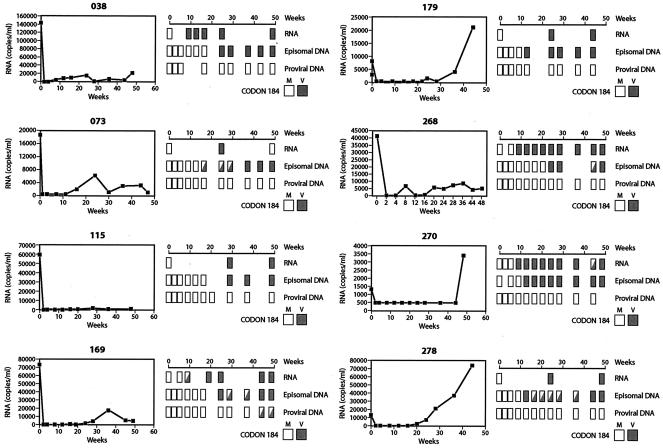
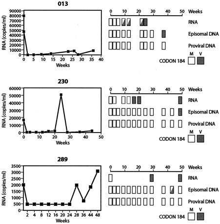
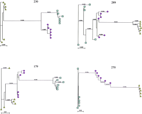
Current regimens for the management of human immunodeficiency virus type 1 (HIV-1) infection suppress plasma viremia to below detectable levels for prolonged intervals. Nevertheless, there is a rapid resumption in plasma viremia if therapy is interrupted. Attempts to characterize the extent of viral replication under conditions of potent suppression and undetectable plasma viremia have been hampered by a lack of convenient assays that can distinguish latent from ongoing viral replication. Using episomal viral cDNA as a surrogate for ongoing replication, we previously presented evidence that viral replication persists in the majority of infected individuals with a sustained aviremic status. The labile nature of viral episomes and hence their validity as surrogate markers of ongoing replication in individuals with long-term-suppressed HIV-1 infection have been analyzed in short-term in vitro experiments with conflicting results. Since these in vitro experiments do not shed light on the long-term in vivo dynamics of episomal cDNA or recapitulate the natural targets of infection in vivo, we have analyzed the dynamics of episomal cDNA turnover in vivo by following the emergence of an M184V polymorphism in plasma viral RNA, in episomal cDNA, and in proviral DNA in patients on suboptimal therapies. We demonstrate that during acquisition of drug resistance, wild-type episomal cDNAs are replaced by M184V-harboring episomes. Importantly, a complete replacement of wild-type episomes with M184V-containing episomes occurred while proviruses remained wild type. This indicates that episomal cDNAs are turned over by degradation rather than through death or tissue redistribution of the infected cell itself. Therefore, evolution of episomal viral cDNAs is a valid surrogate of ongoing viral replication in HIV-1-infected individuals.


Figures

FIG. 1.
FIG. 1.
Strategy for long-range PCR amplification of episomal HIV-1 cDNAs containing RT or envelope sequences. Major cDNA intermediates in reverse transcription are denoted. Thin line, viral RNA; thick line, cDNA. Primer-binding sites for initiation of minus-strand cDNA synthesis and polypurine tracks for plus- or minus-strand synthesis are indicated by open circles and squares, respectively. First-, second-, and third-round primer sets are indicated next to the specific cDNA intermediate they are designed to amplify. HIV-1 pol sequences were amplified using Jumpstart REDAccuTaq DNA polymerase (Sigma), following the manufacturer's protocol with the following modifications. Amplifications were done in a 50-μl reaction mixture containing 1× buffer, 0.5 mM deoxynucleoside triphosphates, 0.5 μM primers, 1.5 U of REDAccuTaq, and 10 μl (10% of total) of either episomal or chromosomal DNA purified from patient peripheral blood lympocytes. After an initial denaturation at 94°C for 2 min, first-round products were generated by cycling 25 to 30 times at 94°C for 15 s, 60°C for 30 s, and 68°C for 1 min per kilobase pair of target sequence. The first-round extension time for Alu PCR was 5 min. One or two microliters from the first round was then subjected to 20 to 30 cycles of amplification under the same reaction conditions.
FIG. 2.
FIG. 2.
Evolution of codon 184 of HIV-1 RT in episomal and proviral cDNAs in patients on ZDV-3TC therapy. For each patient, plasma viral RNA loads were monitored after the initiation (week 0) of dual therapy. In each case, an M-to-V substitution at codon 184, which confers resistance to 3TC, was evident at week 24 post-therapy initiation. At various intervals following initiation of ZDV-3TC therapy, the amino acid identity for codon 184 was determined in plasma viral RNA, episomal DNA, and proviral DNA. Boxes indicate the sampling intervals. A solid box denotes the exclusive presence of the M184V mutation in sequence electropherograms of PCR products at that sampling interval. Partially filled boxes denote the presence of both wild-type and M184V genotypes at that sampling interval.
FIG. 3.
FIG. 3.
Delayed emergence of M184V mutations in episomal cDNA in patients on dual nucleoside therapy. Symbols are as described in the legend for Fig. 2.
FIG. 4.
FIG. 4.
Phylogenetic relationships in episomal (2-LTR circle) envelope genes in patients with rapid and delayed evolution of M184V polymorphisms in episomal cDNA. Nested PCR was used to specifically amplify envelope sequences from 2-LTR circles, using primers detailed in Table 1. Nucleotide sequences corresponding to the C2 and V3 regions of the envelope glycoprotein were aligned by using Clustal W (MacVector 7.1.1), and the phylogenetic relationships were generated by using the neighbor joining method included in the Mac Vector software package with a Kimura-2 parameters distance matrix. Six unique nucleotide sequences spanning C2 and V3 were determined at weeks 0 (circles), 24 (squares), and 48 (triangles) of the study for two patients (179 and 270) with rapid emergence of M184V episomes and two patients (230 and 289) with delayed emergence of the M184V polymorphism in episomal cDNA. Median genetic distances for early and late converters were significantly different (P < 0.03) in a Mann-Whitney nonparametric analysis.

References

    1. Bi, X., H. Gatanaga, S. Ida, K. Tsuchiya, S. Matsuoka-Aizawa, S. Kimura, and S. Oka. 2003. Emergence of protease inhibitor resistance-associated mutations in plasma HIV-1 precedes that in proviruses of peripheral blood mononuclear cells by more than a year. J. Acquir. Immune Defic. Syndr. 34:1-6. - PubMed
    1. Brooks, D. G., D. H. Hamer, P. A. Arlen, L. Gao, G. Bristol, C. M. Kitchen, E. A. Berger, and J. A. Zack. 2003. Molecular characterization, reactivation, and depletion of latent HIV. Immunity 19:413-423. - PubMed
    1. Bushman, F. 2003. Measuring covert HIV replication during HAART: the abundance of 2-LTR circles is not a reliable marker. AIDS 17:749-750. - PubMed
    1. Butler, S. L., E. P. Johnson, and F. D. Bushman. 2002. Human immunodeficiency virus cDNA metabolism: notable stability of two long-terminal repeat circles. J. Virol. 76:3739-3747. - PMC - PubMed
    1. Chun, T. W., L. Carruth, D. Finzi, X. Shen, J. A. DiGiuseppe, H. Taylor, M. Hermankova, K. Chadwick, J. Margolick, T. C. Quinn, Y. H. Kuo, R. Brookmeyer, M. A. Zeiger, P. Barditch-Crovo, and R. F. Siliciano. 1997. Quantification of latent tissue reservoirs and total body viral load in HIV-1 infection. Nature 387:183-188. - PubMed

Publication types

MeSH terms

LinkOut - more resources