. 2005 Mar 28;168(7):999-1012.
doi: 10.1083/jcb.200412076.

The yeast S phase checkpoint enables replicating chromosomes to bi-orient and restrain spindle extension during S phase distress

Affiliations

The yeast S phase checkpoint enables replicating chromosomes to bi-orient and restrain spindle extension during S phase distress

Jeff Bachant et al. J Cell Biol. .

Abstract

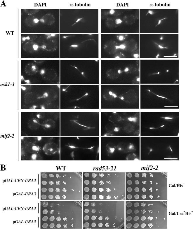
The budding yeast S phase checkpoint responds to hydroxyurea-induced nucleotide depletion by preventing replication fork collapse and the segregation of unreplicated chromosomes. Although the block to chromosome segregation has been thought to occur by inhibiting anaphase, we show checkpoint-defective rad53 mutants undergo cycles of spindle extension and collapse after hydroxyurea treatment that are distinct from anaphase cells. Furthermore, chromatid cohesion, whose dissolution triggers anaphase, is dispensable for S phase checkpoint arrest. Kinetochore-spindle attachments are required to prevent spindle extension during replication blocks, and chromosomes with two centromeres or an origin of replication juxtaposed to a centromere rescue the rad53 checkpoint defect. These observations suggest that checkpoint signaling is required to generate an inward force involved in maintaining preanaphase spindle integrity during DNA replication distress. We propose that by promoting replication fork integrity under these conditions Rad53 ensures centromere duplication. Replicating chromosomes can then bi-orient in a cohesin-independent manner to restrain untimely spindle extension.


Figures

Figure 1.
Figure 1.
Spindle extension in HU-treated rad53-21 mutants. (A) WT (JBY430) and rad53-21 (JBY1201) GFP-TUB1 TRP1-GFP cells were released from G1 into 200 mM HU and visualized by GFP fluorescence and low intensity bright-field illumination after 90 min. Arrows,TRP1-GFP foci; pointers, reduced tubulin-GFP in rad53 spindles. Bar, 5 μm. (B) Pseudocolored images of chromatin (DAPI; red) and spindle poles (SPC42-GFP; green) in HU-treated rad53-21 mutants (JBY1274) 2 h after release from G1 into 200 mM HU. Bar, 5 μm. (C) Spindle length in HU-arrested WT (JBY1129) and rad53-21 (JBY1274) SPC42-GFP cells. The distance between Spc42-GFP foci was measured in 500 cells 2 h after release from G1 into 200 mM HU. The percentage of spindles ≥3 μm is indicated. (D) Spindle extension kinetics in WT (Y300) and rad53-21 (Y301) strains. Time points from cultures released from G1 with (right) or without (left) 200 mM HU were processed for FACS and α-tubulin immunofluorescence. The percentage of spindles ≥3 μm (open squares, WT; open circles, rad53-21) and budded cells (hatched square, WT; hatched circle, rad53-21) was determined. (E) Clb2-Cdk1 activity in WT (JBY012; left) and rad53-21 (JBY013; right) cells. Cells harboring Clb2-HA were released from G1 in the presence (circles) or absence (squares) of 200 mM HU, and histone H1 kinase activity in α-HA immunoprecipitates was quantified.
Figure 2.
Figure 2.
Spindle dynamics in rad53-21 mutants. WT (JBY1129) and rad53-21 (JBY1274) SPC42-GFP strains were released from G1 into 200 mM HU. Cells were prepared for imaging 75–90 min after release. Stacks of Spc42-GFP images were acquired every 2 min. Numbers indicate lapsed time. (A) 20-min time-lapse sequence for a WT cell. (B and C) SPB extension and collapse in two HU-treated rad53-21 cells. (D) Graphs depicting changes in spindle length over time for four WT and three rad53-21 cells.
Figure 3.
Figure 3.
SCC1 is not required to prevent spindle extension during HU arrest. WT (Y300), rad53-21 (Y301), and scc1-73 (JBY585) strains were released from G1 into 200 mM HU at 35°C. cdc23-1 (JBY622), cdc23-1rad53-21 (JBY623), and cdc23-1scc1-73 (JBY1305) strains were released at 35°C in the absence of HU. (A) Spindle (α-tubulin) and chromosome (DAPI) morphology in HU- or cdc23-arrested strains 2.5 h after G1 release. Bars, 5 μm. (B) Kinetics of spindle extension and budding. (left) HU-arrested strains (squares, WT; circles, rad53-21; triangles, scc1-73). (right) cdc23-1–arrested strains (squares, cdc23-1; circles, cdc23-1rad53-21; triangles, cdc23-1scc1-73).
Figure 4.
Figure 4.
Analysis of Esh mutants. (A) Cell survival after HU treatment. Cultures of WT (Y300; squares), smt4-3 (JBY047; triangles), ask1-1 (JBY368; circles), and rad53-21 (Y301; diamonds) cells were exposed to 200 mM HU. At the indicated times, aliquots were plated for cell viability. (B) Spindle length during HU arrest. The distance between Spc42-GFP foci was measured for 250 WT (JBY1129), smt4-3 (JBY1312), and ask1-1 (JBY1196) SPC42-GFP cells 2.5 h after release from G1 into 200 mM HU. The percentage of spindles ≥3 μm is indicated. (C) MIF2 clones isolated as Smt4 two-hybrid interactors. Regions of similarity to CENP-C (black boxes) and the HMGI (Y) domain (gray box) are indicated. pDAB (GAL4 DNA binding domain [DBD] vector), pACT (GAL4 activation domain [AD] vector), pSE1111 (SNF4-DBD), pJBN84 (SMT4-DBD), and MIF2-AD clones p20.1, p53.1, and p59.1 were analyzed in the indicated combinations. Growth on TrpLeuAde media indicates a two-hybrid interaction.
Figure 5.
Figure 5.
MIF2 and ASK1 restrain spindle extension during HU arrest. (A) Cell survival in HU-treated mif2-2 mutants. Cultures of WT (Y300) and mif2-2 (JBY358) strains were treated ± 200 mM HU at 24 or 35°C. Aliquots were plated to monitor cell viability. Squares, WT/24°C/HU; triangles, WT/35°C/HU; diamonds, mif2-2/24°C; circles, mif2-2/24°C/HU; hatched diamonds, mif2-2/35°C/HU; hatched squares, mif2-2/35°C. (B) Spindle length was measured for 250 WT SPC42-GFP (JBY1129), ask1-3 SPC42-GFP (JBY1325), and mif2-2 (JBY358) cells 2.5 h after G1 release into 200 mM HU. The percentage of spindles ≥3 μm is indicated. (C) Kinetics of spindle extension during HU arrest. WT (Y300, squares), rad53-21 (Y301; diamonds), mif2-2 (JBY358; triangles), and ask1-3 (JBY1325; circles) were released from G1 into 200 mM HU at 35°C. Time points were processed for DAPI and α-tubulin staining.
Figure 6.
Figure 6.
Analysis of spindle morphology in HU-treated ask1 and mif2 mutants and CEN function in rad53 mutants. (A) Spindle extension after activation of the S phase checkpoint. 2.5 h after release from G1 to 200 mM HU at 35°C, DAPI and α-tubulin images were obtained for WT, ask1-3, and mif2-2 strains. Bars, 5 μm. (B) CEN transcription assay. 10-fold serial dilutions of WT (Y300), rad53-21 (Y301), and mif2-2 (JBY358) strains transformed with pGAL-CEN6-URA3 or pGAL-URA3 were spotted onto Gal/His and Gal/HisUra galactose media. Growth was assayed after 4 d at 28°C. Growth on His media selects for the plasmid; HisUra plates reveal the extent of CEN interference with URA3 expression.
Figure 7.
Figure 7.
Spindle extension in KT mutants during HU or metaphase arrest. cdc23-1 (JBY1289), cdc23-1rad53-21 (JBY1293), cdc23-1ndc10-1 (JBY1367), cdc23-1ndc80-1 (JBY1363), cdc23-1dam1-1 (JBY1333), and cdc23-1ipl1-321 (JBY1357) SPC42-GFP strains were released from G1 ± 200 mM HU at 35°C. After 2.5 h, the distance between Spc42-GFP foci was measured for 250 cells. The percentage of spindles ≥4 μm (−HU) or ≥3 μm (+HU) is depicted.
Figure 8.
Figure 8.
HU-treated mad2 and ipl1 mutants. (A) Spindle length was measured in 250 WT (Y300), rad53-21 (Y301), mad2-Δ (JBY1393), and rad53-21mad2-Δ (JBY1395) cells 2.5 h after release from G1 into 200 mM HU. The percentage of spindles ≥3 μm is indicated. (B) Pseudocolored images of chromatin (DAPI; red) and spindle poles (Spc42-GFP; green) in HU-treated WT (JBY1129), rad53-21 (JBY1274), ipl1-321 (JBY1353), and ipl1-321rad53-21 (JBY1389) SPC42-GFP strains 2.5 h after G1 release into 200 mM HU. Bar, 5 μm.
Figure 9.
Figure 9.
Dicentric bridging restricts spindle extension in HU-treated rad53 mutants. LEU2-GFP (JBY541), his4::GAL-CEN3 LEU2-GFP (JBY1208), rad53-21 LEU2-GFP (JBY1201), and two rad53-21 his4::GAL-CEN3 LEU2-GFP segregants (JBY1203 and JBY1206) were propagated in galactose media, placed at G1 arrest, and released into 200 mM HU glucose media to activate the dicentric. (A) After 2 h, cells were processed for α-tubulin immunofluorescence and spindle length was measured for 250 cells. The percentage of spindles ≥3 μm is indicated. (B) Starting at 90 min after release, GFP-tagged chromatin between the dicentric CENs was visualized by fluorescence. Bars, 5 μm.
Figure 10.
Figure 10.
pCENARS minichromosomes suppress spindle extension in HU-treated rad53 mutants. WT (JBY1129) and rad53-21 (JBY1274) SPC42-GFP-TRP1 strains were transformed with one (pRS413), two (pRS413 and pRS415), or three (pRS413, pRS415, and pRS416) pCENARS plasmids (Sikorski and Hieter, 1989). WT (Y300) and rad53-21 (Y301) were transformed with four pCENARS plasmids (pRS413, pRS414, pRS415, and pRS416). Transformants were cultured to maintain the plasmids, arrested in G1, and released into yeast extract/peptone/dextrose media containing 200 mM HU. Spindle length was measured in 200 cells at the indicated times using Spc42-GFP foci or α-tubulin immunofluorescence. (A) Effect of pCENARS dosage on spindle extension in HU-treated rad53 mutants (right). (left) Budding kinetics. (B) Spindle length distribution in WT and rad53 mutants after 2.5 h of HU treatment. The percentage of spindles ≥3 μm is indicated. (C) Requirements for pCENARS suppression. WT (JBY1129), rad53-21 (JBY1274), ndc80-1 (JBY1359), and ndc80-1rad53-21 (JBY1400) SPC42-GFP strains and dbf4-1 (JBY997), dbf4-1rad53-21 (JBY1001), mad2-Δ (JBY1393), mad2-Δrad53-21 (JBY1395), scc1-73 (JBY585), and scc1-73rad53-21 (JBY1397) strains ± three pCENARS plasmids were released from G1 into 200 mM HU at 35°C. After 2.5 h, the percentage of spindles ≥3 μm was determined for 200 cells.

References

    1. Agarwal, R., Z. Tang, H. Yu, and O. Cohen-Fix. 2003. Two distinct pathways for inhibiting Pds1 ubiquitination in response to DNA damage. J. Biol. Chem. 278:45027–45033. - PubMed
    1. Alcasabas, A., A. Osborn, J. Bachant, F. Hu, P. Werler, K. Bousset, K. Kanji-Furuya, J.F.X. Diffley, A. Carr, and S.J. Elledge. 2001. Mrc1 transduces DNA replication stress signals to activate Rad53. Nat. Cell Biol. 3:958–965. - PubMed
    1. Allen, J.B., Z. Zhou, W. Siede, E.C. Friedberg, and S.J. Elledge. 1994. The SAD1/RAD53 protein kinase controls multiple checkpoints and DNA damage-induced transcription in yeast. Genes Dev. 8:2401–2415. - PubMed
    1. Bachant, J., A. Alcasabas, Y. Blat, N. Kleckner, and S.J. Elledge. 2002. The SUMO-1 isopeptidase Smt4 is linked to centromeric cohesion through SUMO-1 modification of DNA topoisomerase II. Mol. Cell. 9:1169–1182. - PubMed
    1. Biggins, S., and A.W. Murray. 2001. The budding yeast protein kinase Ipl1/Aurora allows the absence of tension to activate the spindle checkpoint. Genes Dev. 15:3118–3129. - PMC - PubMed

Publication types

MeSH terms