Comparative Study
. 2005 Jun;95(7):1113-30.
doi: 10.1093/aob/mci134. Epub 2005 Mar 29.

Comparative structure and pollen production of the stamens and pollinator-deceptive staminodes of Commelina coelestis and C. dianthifolia (Commelinaceae)

Affiliations
Comparative Study

Comparative structure and pollen production of the stamens and pollinator-deceptive staminodes of Commelina coelestis and C. dianthifolia (Commelinaceae)

William C Hrycan et al. Ann Bot. 2005 Jun.

Abstract

Background and aims: Flowers of Commelina coelestis and C. dianthifolia provide pollen alone as a floral reward, and rely on visual cues to attract pollinators. Three stamen types, all producing pollen, occur in each of these species: two cryptically coloured lateral stamens, a single cryptically coloured central stamen and three bright yellow staminodes that sharply contrast with the blue to purple corolla. The objective was to compare the stamen structure and pollen characteristics of each of the three stamen types, and to test the hypothesis that the staminodes are poor contributors of viable pollen for the siring of seed. The pollination roles of the three stamen types and the breeding systems of both species were also explored.

Methods: Light, fluorescence and scanning electron microscopy were utilized to examine stamen morphology and pollen structure and viability. Controlled hand pollinations were used to explore the breeding system of each species. Filament and style lengths were measured to investigate herkogamy and autogamy.

Key results: Pollen from all stamen morphs is viable, but staminode pollen has significantly lower viability. Pollen polymorphism exists both (a) between the lateral and central stamens and the staminodes, and (b) within each anther. Lateral and central stamens have thicker endothecia with a greater number of secondary cell wall thickenings than the staminodes.

Conclusions: Both species are entomophilous and facultatively autogamous. Lateral stamen pollen is important for cross-pollination, central stamen pollen is utilized by both species as a pollinator reward and for delayed autogamy in C. dianthifolia, and the staminodes mimic, by means of both colour and epidermal features, large amounts of pollen to attract insects to the flowers. Pollen from all three anther morphs is capable of siring seed, although staminode pollen is inferior. The thin staminode endothecium with fewer secondary thickenings retards staminode dehiscence.


Figures

F<sc>ig</sc>. 1.
Fig. 1.
Flowers of Commelina coelestis (A) and C. dianthifolia (B), shortly after anthesis.
F<sc>ig</sc>. 2.
Fig. 2.
Whole flowers and anther macrophotography of C. coelestis (B, C, E, G and I) and C. dianthifolia (A, D, F and H). (A) Transverse section showing arrangement of lateral stamens (ls), central stamen (cs), staminodes (st), ovary (ov), petals (p) and sepals (s). Arrowheads indicate ovary trichomes. Scale bar = 0·5 mm. (B) Transverse section of flower showing arrangement of lateral stamens (ls), central stamen (cs) and staminodes (st). Arrowheads indicate ovary trichomes. Scale bar = 0·5 mm. (C) Inflorescence with petals and sepals of open flower removed. Lateral stamens (ls), central stamen (cs) and staminodes (st). Maturing buds at right. (D) SEM of whole flower with petals and sepals removed. Lateral stamens (ls), central stamen (cs), staminodes (st), stigma (sg) and style (sy). Trichomes (arrowheads) are visible on the ovary wall. Scale bar = 1 mm. (E) Adaxial (left) and abaxial (right) view of dehisced lateral stamens showing abundant yellow pollen. (F) Abaxial view of lateral stamen. (G and H) Central stamen at dehiscence. Note patches of purple and white. (I) Two of three staminodes from a single flower in abaxial (left) and adaxial (right) view showing arrangement of sterile lobes (sl) and fertile lobes (fl).
F<sc>ig</sc>. 3.
Fig. 3.
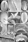
SEM of lateral (A–F) and central (G–K) stamens of C. coelestis (B, D, E, I and J) and C. dianthifolia (A, C, F–H and K). (A) Adaxial side of pre-dehiscent anther and connective tissue between thecae. Scale bar = 0·25 mm. (B) Stomium of pre-dehiscent anther beginning to open (arrowhead). Scale bar = 0·1 mm. (C) Adaxial side of dehisced lateral stamen. Reflexed anther walls, residual pollen grains (po) and remnants of septum (se) are evident. Scale bar = 0·25 mm. (D) Abaxial side of pre-dehiscent anther. Scale bar = 0·25 mm. (E) Apical view of dehiscent anther. Scale bar = 0·25 mm. (F) Abaxial side of dehiscent anther showing reflexed walls. Scale bar = 0·25 mm. (G) Pre-dehiscent, curved anther. Scale bar = 0·25 mm. (H) Abaxial view of pre-dehiscent anther. Scale bar = 0·25 mm. (I) Lip cells (lc) at stomium. Scale bar = 50 μm. (J) Side view of dehisced anther. Scale bar = 0·25 mm. (K) Side view of fully dehisced anther, showing reflexed walls centrally, residual pollen grains (po), and remnants of septum (se). Scale bar = 0·25 mm.
F<sc>ig</sc>. 4.
Fig. 4.
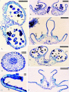
Paraffin sections of lateral (A–C) and central (D–G) stamens of C. coelestis (B, D and G) and C. dianthifolia (A, C, E and F). (A) Half of pre-dehiscent anther in transverse section with locules (lo) separated by intact septum (se); endothecium (en) and epidermis (ep). Scale bar = 0·1 mm. (B) Dehisced anther with cavity (ca) in connective tissue. Walls reflexing. Filament (ft). Scale bar = 0·25 mm. (C) Fully dehisced anther in transverse section with residual pollen (po) and remnants of septa (se). Cavities (ca) in connective tissue are visible interior to both septa. Section of stigma (sg) shows stigmatic papillae (sp) and adhering pollen grains (po). Scale bar = 0·25 mm. (D) Filament of central stamen with single vascular bundle. Scale bar = 50 μm. (E) Pre-dehiscent anther with ruptured stomium (sm), partially degraded septa (se), and connective tissue cavities (ca) bordering vascular bundle. Scale bar = 0·1 mm. (F) Fully dehisced anther with connective tissue cavities (ca). Upper walls reflexed whereas lower walls maintain horizontal position. Curled anther wall of adjacent lateral stamen at bottom right. Scale bar = 0·25 mm. (G) Curled upper wall of anther. Note endothecium (en) and epidermis (ep). Scale bar = 10 μm.
F<sc>ig</sc>. 5.
Fig. 5.
SEM of staminodes of C. coelestis (A, B and E–H) and C. dianthifolia (C and D). (A and B) Staminodes showing the variation present between staminodes within a flower. Upper and lower lobes are sterile (sl) and the central lobes are fertile (fl). Scale bars = 0·25 mm. (C) Newly dehiscent and (D) fully dehiscent staminodes. Scale bars = 0·25 mm. (E) Pre-dehiscent and (F) post-dehisced fertile lobes. Reflexed theca walls expose pollen (po) within. (G) Surface of sterile lobe with isodiametric papillae. Scale bar = 10 μm. (H) Higher magnification of sterile lobe showing surface detail. Scale bar = 10 μm.
F<sc>ig</sc>. 6.
Fig. 6.
Paraffin sections of staminodes of C. coelestis (A and C–G) and C. dianthifolia (B and E). (A) Longitudinal section showing four sterile lobes of connective tissue (sl) around two fertile thecae (fl). Undulating filament (ft) with solitary vascular bundle. Scale bar = 0·25 mm. (B) Transverse section of staminode filament with single vascular bundle. Scale bar = 0·1 mm. (C) Longitudinal section of fertile theca with pollen (po) in locule (lo) lined by endothecium (en). Scale bar = 0·1 mm. (D) Near tangential section of layer of endothecium (en) lining the same locule (lo). Scale bar = 0·1 mm. (E) Transverse sections through fertile lobes of dehisced staminode of C. coelestis (bottom) and pre-dehiscent staminode of C. dianthifolia (top). Scale bar = 0·5 mm. (F) Transverse section of dehisced fertile lobe. Scale bar = 0·1 mm. (G) Close-up of anther wall in transverse section, showing endothecium (en) below nucleated epidermis (ep). Scale bar = 25 μm.
F<sc>ig</sc>. 7.
Fig. 7.
Pollen and features of the gynoecium of C. coelestis (A, E, G, H and K–M) and C. dianthifolia (B–D, F, I and J). (A and B) Dimorphic pollen of central stamen. Scale bars = 50 μm. (C) Dimorphic pollen of staminode. Scale bar = 50 μm. (D) SEM of dimorphic pollen of lateral stamen. Scale bar = 10 μm. (E) Large pollen grain of central stamen, treated with OsO4 vapour. Scale bar = 10 μm. (F) Small pollen grain inside locule of central stamen. Secondary thickenings (arrowheads) are visible through primary wall of endothecium (en) lining locule. Scale bar = 10 μm. (G) Large pollen grain of staminode with adhering raphide crystals (r). Treated with OsO4 vapour. Scale bar = 10 μm. (H) Small pollen grain of central stamen with adhering raphide crystals (r). Treated with OsO4 vapour. Scale bar = 10 μm. (I and J) Fluorochromatic reaction (FCR) test on lateral stamen pollen. Large pollen (L), small pollen (S) and intermediately sized pollen (I). (I) Pollen viewed under normal light and (H) viewed under fluorescence. Large pollen grains fluoresced brightly but small and intermediate grains did not fluoresce. Scale bars = 0·1 mm. (K) Autogamy mechanism, with the stigma pushed against the dehisced central stamen. Scale bar = 1 mm. (L) Pollen (left) adhering to three-lobed stigma pushed against anther of central stamen; stigmatic papillae evident at right. Scale bar = 0·1 mm. (M) Glandular trichome on ovary wall. Scale bar = 10 μm.
F<sc>ig</sc>. 8.
Fig. 8.
Change in gynoecial length over time compared with filament lengths of the lateral and central stamens: (A) Commelina coelestis; (B) C. dianthifolia.

Similar articles

Cited by

References

    1. Baker FJ, Silverton RE. 1976.Introduction to medical laboratory technology, 5th edn. London: Butterworths.
    1. Brantjes NBM. 1980. Individualisme bij bloembezoekende insekten op Commelina [Individualism by flower-seeking insects on Commelina]. De Levende Natuur 82: 9–16.
    1. Brashier CK. 1966. A revision of Commelina (Plum.) L. in the U.S.A. Bulletin of the Torrey Botanical Club 93: 1–19.
    1. Brewbaker JL, Kwack BH. 1963. The essential role of calcium ion in pollen germination and pollen tube growth. American Journal of Botany 50: 859–865.
    1. Christensen KI, Hansen HV. 1998. SEM-studies of epidermal patterns of petals in the angiosperms. Opera Botanica 135: 1–91.

Publication types