. 2005 Apr;25(8):3007-18.
doi: 10.1128/MCB.25.8.3007-3018.2005.

Identification of a novel, intraperoxisomal pex14-binding site in pex13: association of pex13 with the docking complex is essential for peroxisomal matrix protein import

Affiliations

Identification of a novel, intraperoxisomal pex14-binding site in pex13: association of pex13 with the docking complex is essential for peroxisomal matrix protein import

Annette Schell-Steven et al. Mol Cell Biol. 2005 Apr.

Abstract

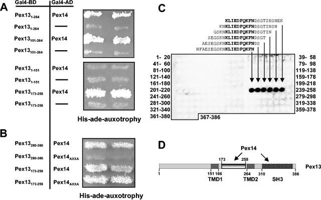
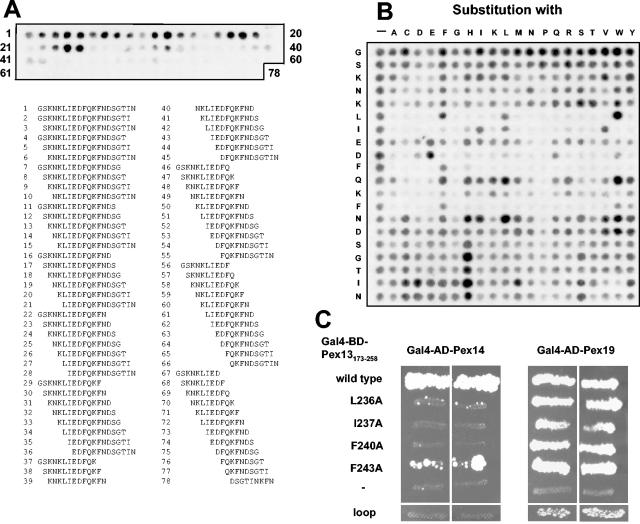
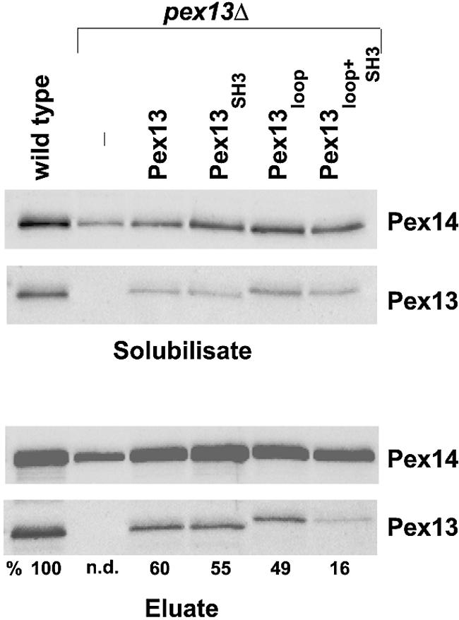
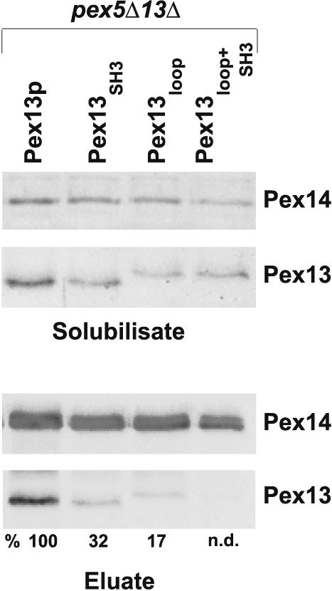
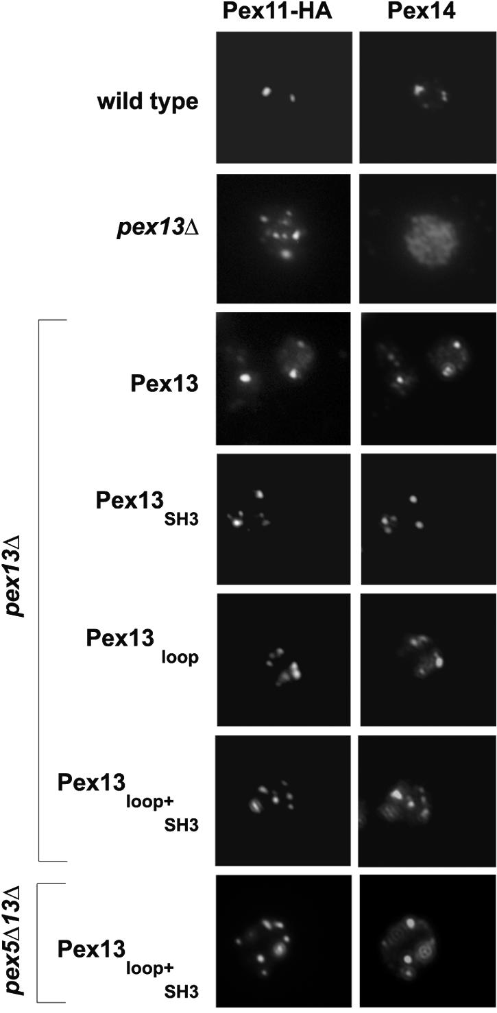
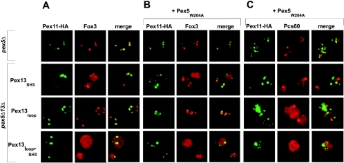
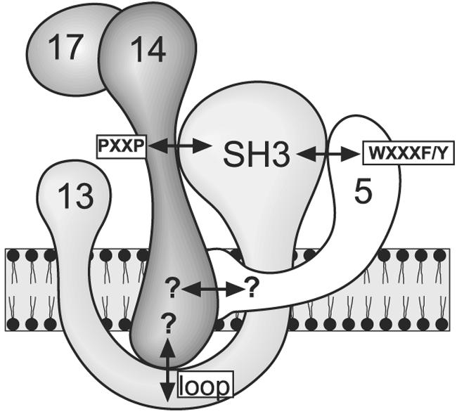
The peroxisomal docking complex is a key component of the import machinery for matrix proteins. The core protein of this complex, Pex14, is thought to represent the initial docking site for the import receptors Pex5 and Pex7. Associated with this complex is a fraction of Pex13, another essential component of the import machinery. Here we demonstrate that Pex13 directly binds Pex14 not only via its SH3 domain but also via a novel intraperoxisomal site. Furthermore, we demonstrate that Pex5 also contributes to the association of Pex13 with Pex14. Peroxisome function was affected only mildly by mutations within the novel Pex14 interaction site of Pex13 or by the non-Pex13-interacting mutant Pex5(W204A). However, when these constructs were tested in combination, PTS1-dependent import and growth on oleic acid were severely compromised. When the SH3 domain-mediated interaction of Pex13 with Pex14 was blocked on top of that, PTS2-dependent matrix protein import was completely compromised and Pex13 was no longer copurified with the docking complex. We conclude that the association of Pex13 with Pex14 is an essential step in peroxisomal protein import that is enabled by two direct interactions and by one that is mediated by Pex5, a result which indicates a novel, receptor-independent function of Pex5.


Figures

FIG. 1.
FIG. 1.
Identification of a novel Pex14-binding site in Pex13. (A) Interactions of Pex13 fragments with Pex14 in a yeast two-hybrid assay. Truncations of PEX13 were fused to the GAL4 DNA-binding domain (Gal4-BD) and coexpressed with a PEX14-GAL4 activation domain (Gal4-AD) fusion in yeast strain PJ69-4A. As control, the bare Gal4-AD was coexpressed with the Pex13-Gal4-BD fusion. Two independent transformants were tested for prototrophy on plates lacking both histidine and adenine. The following plasmids were used to express the indicated Pex13 fragments: Pex131-151 (pKat31), Pex131-264 (pKat33), Pex13151-264 (pKat129), and Pex13173-258 (pKat145). (B) Involvement of the proline-rich motif of Pex14 in binding to Pex13. The two Pex14-interacting fragments of Pex13, comprising the SH3 domain (Pex13280-386) and the novel binding site (Pex13173-258), were tested for interactions with Pex14 mutated in its proline-rich motif (Pex14AXXA; pWG14/6) as described for panel A. (C) In vitro binding of Pex14 to the novel binding site of Pex13. Synthetic 20-mer peptides with two-amino-acid shifts between neighboring peptides and representing full-length Pex13 were synthesized on cellulose membranes. The identities of the first and the last peptides in each line of the peptide array are indicated. The membranes were incubated with purified recombinant GST-Pex14 followed by monoclonal anti-GST antibodies. Pex13 peptides that bound to GST-Pex14p were visualized with horseradish peroxidase-conjugated anti-mouse antibodies and ECL reagent. The sequences of the interacting peptides are shown, and the overlapping amino acids are highlighted by bold type. (D) Schematic view of Pex13 and, its proposed transmembrane domains TMD1 and TMD2, the SH3 domain, and the novel Pex14-binding site. Numbers denote amino acid positions.
FIG. 2.
FIG. 2.
Characterization of the second Pex14-binding site in Pex13. (A) Length analysis of the novel Pex14-binding site. Peptides comprising systematic truncations of Pex14-interacting peptide Pex13231-250 down to a length of nine amino acids were synthesized on cellulose membranes and incubated with purified His6-Pex14. Bound His6-Pex14 was visualized immunologically with monoclonal anti-His6 antibodies. The numbered peptide sequences shown below the membrane correlate with the spot numbers on the membrane. (B) Substitution analysis. Pex14 was tested for interactions with mutated variants of Pex13231-250 peptide GSKNKLIEDFQKFNDSGTIN as described for panel A. The first row represents the nonmutated wild-type peptide, whereas peptides in all other rows harbor the indicated single amino acid substitutions. Spots with reduced intensities represent peptides with reduced binding affinities for Pex14. (C) In vivo effect of mutating critical residues of the Pex14-binding site. (Left panels) A yeast two-hybrid assay was used to study the interaction of Pex14 with Pex13173-258 that had been mutated to A at position L236 (pAS40), I237 (pAS41), F240 (pAS42), or F243 (pAS43) or at all four positions (loop; pAS82). (Right panels) As a control, the Pex13 fragments were also assayed for interactions with Pex19, which require amino acids 200 to 220 of Pex13. BD, DNA-binding domain; AD, activation domain.
FIG. 3.
FIG. 3.
Association of Pex13 mutant proteins with the docking complex. The indicated oleic acid-induced strains containing a chromosomal protein A tag at the PEX14 locus were analyzed for the presence of Pex13 in the docking complex that had been purified with ProtA-Pex14. In particular, the UTL-7A-derived pex13Δ mutant strains harboring plasmids designed to express Pex13 (pAS62), Pex13loop (pAS76), Pex13SH3 (pAS61), and Pex13loop+SH3 (pAS75) were analyzed. (Upper panels) Immunoblots loaded with aliquots of proteins solubilized with 1% digitonin from total membrane fractions and decorated with anti-Pex13 and Pex14 antibodies. (Lower panels) Immunoblots showing the abundance of Pex14 and Pex13 in eluates. Numbers below the bottom panel denote the ratios of the intensities of the Pex13 signals to the corresponding Pex14 signals. The calculated intensity of the wild-type sample was set at 100%. n.d., not determined.
FIG. 4.
FIG. 4.
Dependence of peroxisomal matrix protein import on direct Pex13-Pex14 interactions. Oleic acid-induced wild-type and pex13Δ cells expressing HA-Pex11 were subjected to double-immunofluorescence microscopy so as to localize HA-Pex11 in tandem with PTS1 protein Pcs60 (A), PTS2 protein Fox3 (B), or PTS1-like protein Cta1 (C). The same procedure was applied to pex13Δ cells expressing Pex13 (pKat113), Pex13SH3 (pAS53), Pex13loop (pAS71), and Pex13loop+SH3 (pAS73). Detection was achieved with mouse monoclonal antibodies against the HA epitope combined with rabbit polyclonal antibodies against the individual matrix proteins. As secondary antibodies, Alexa Fluor 488-labeled anti-mouse IgG and Alexa Fluor 594-labeled anti-rabbit IgG were used. A congruent fluorescence pattern denotes colocalization of HA-Pex11 with the analyzed matrix proteins, a finding which is readily revealed in the merged images. (D) From the same cells, a postnuclear supernatant (PNS) was produced and subfractionated into a 25,000 × g pellet fraction (P) enriched for peroxisomes and a supernatant fraction (SN) enriched for the cytosol. Equivalent volumes of fractions were loaded on the gels, transferred to nitrocellulose membranes, and analyzed for the distributions of the specified proteins.
FIG. 5.
FIG. 5.
Involvement of Pex5 in the association of Pex13 with the Pex14-containing docking complex. The docking complex was isolated with ProtA-Pex14 from oleic acid-induced cells lacking Pex5 and expressing the indicated mutant versions of Pex13. (Lower panels) The amount of Pex13 coeluting with the docking complex was determined by immunoblot analysis. Numbers below the bottom panel denote the ratios of the intensities of the Pex13 signals to the corresponding Pex14 signals. The calculated intensity of the sample derived from pex5Δ pex13Δ cells expressing wild-type Pex13 was set at 100%. n.d., not determined. (Upper panels) Amounts of Pex14 and Pex13 present in solubilized membrane protein fractions.
FIG. 6.
FIG. 6.
Influence of mutant versions of Pex13 and of Pex5 on Pex14 targeting. The localization of Pex14 in oleic acid-induced pex13Δ and pex5Δ pex13Δ cells expressing wild-type or mutant versions of Pex13 was compared to that of HA-Pex11 by double-immunofluorescence microscopy. Detection was achieved with rabbit polyclonal antibodies against Pex14 and anti-HA monoclonal antibodies followed by Alexa Fluor 488-labeled anti-mouse and Alexa Fluor 594-labeled anti-rabbit secondary antibodies.
FIG. 7.
FIG. 7.
Dependence of matrix protein import on Pex5 in the absence of direct Pex13-Pex14 interactions. (A) Oleic acid-induced pex5Δ pex13Δ cells expressing mutant versions of Pex13 in combination with HA-Pex11 were processed for double-immunofluorescence microscopy with rabbit antibodies against PTS2 protein Fox3 and anti-HA monoclonal antibodies. The secondary antibodies used were Alexa Fluor 488-labeled anti-mouse IgG and Alexa Fluor 594-labeled anti-rabbit IgG. pex5Δ cells were used as a control for intact PTS2 import. (B and C) The same cells but additionally expressing Pex5W204A were similarly inspected for the localization of Fox3 (B) and Pcs60 (C).
FIG. 8.
FIG. 8.
Effect of weakening the Pex13-Pex14 association on peroxisome function. Complementation analysis of pex13Δ cells expressing Pex13, Pex13SH3, Pex13loop, or Pex13loop+SH3 (A) and pex5Δ pex13Δ cells expressing Pex5W204A together with Pex13, Pex13SH3, Pex13ploop, or Pex13loop+SH3 (B) was carried out. The indicated cells were spotted as a series of 10-fold dilutions on oleic acid plates and incubated at 30°C for 5 days.
FIG. 9.
FIG. 9.
Graphic representation of Pex13-Pex14 interactions. Protein-protein interactions between S. cerevisiae peroxins Pex5, Pex13, and Pex14 are denoted by double-headed arrows. Mapped interactions are labeled according to the designations used in this work. Published interactions that had not yet been mapped are indicated by question marks.

Similar articles

Cited by

References

    1. Agne, B., N. M. Meindl, K. Niederhoff, H. Einwachter, P. Rehling, A. Sickmann, H. E. Meyer, W. Girzalsky, and W. H. Kunau. 2003. Pex8p: an intraperoxisomal organizer of the peroxisomal import machinery. Mol. Cell 11:635-646. - PubMed
    1. Albertini, M., P. Rehling, R. Erdmann, W. Girzalsky, J. A. Kiel, M. Veenhuis, and W. H. Kunau. 1997. Pex14p, a peroxisomal membrane protein binding both receptors of the two PTS-dependent import pathways. Cell 89:83-92. - PubMed
    1. Barnett, P., G. Bottger, A. T. Klein, H. F. Tabak, and B. Distel. 2000. The peroxisomal membrane protein Pex13p shows a novel mode of SH3 interaction. EMBO J. 19:6382-6391. - PMC - PubMed
    1. Blobel, F., and R. Erdmann. 1996. Identification of a yeast peroxisomal member of the family of AMP-binding proteins. Eur. J. Biochem. 240:468-476. - PubMed
    1. Bottger, G., P. Barnett, A. T. Klein, A. Kragt, H. F. Tabak, and B. Distel. 2000. Saccharomyces cerevisiae PTS1 receptor Pex5p interacts with the SH3 domain of the peroxisomal membrane protein Pex13p in an unconventional, non-PXXP-related manner. Mol. Biol. Cell 11:3963-3976. - PMC - PubMed

Publication types

MeSH terms

Substances