. 2005 Apr;25(8):3261-75.
doi: 10.1128/MCB.25.8.3261-3275.2005.

Myocyte enhancer factor 2 activates promoter sequences of the human AbetaH-J-J locus, encoding aspartyl-beta-hydroxylase, junctin, and junctate

Affiliations

Myocyte enhancer factor 2 activates promoter sequences of the human AbetaH-J-J locus, encoding aspartyl-beta-hydroxylase, junctin, and junctate

Giordana Feriotto et al. Mol Cell Biol. 2005 Apr.

Abstract

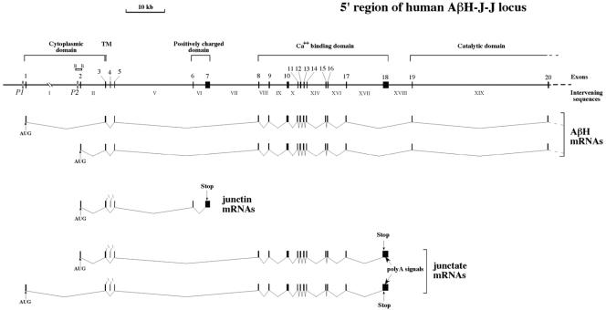
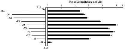
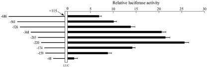
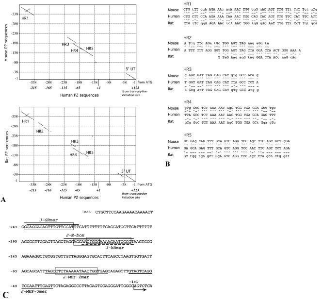
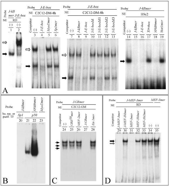
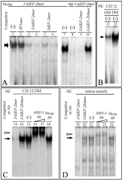
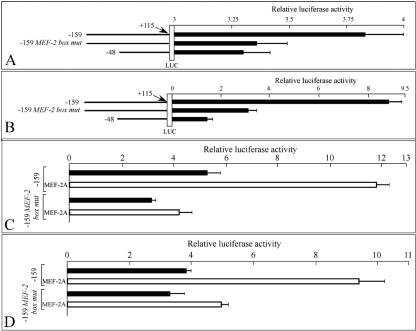
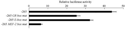
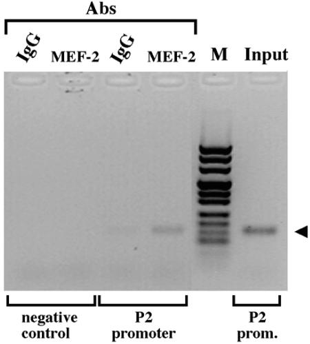
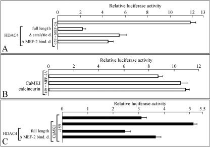
Alternative splicing of the locus AbetaH-J-J generates three functionally distinct proteins: an enzyme, AbetaH (aspartyl-beta-hydroxylase), a structural protein of the sarcoplasmic reticulum membrane (junctin), and an integral membrane calcium binding protein (junctate). Junctin and junctate are two important proteins involved in calcium regulation in eukaryotic cells. To understand the regulation of these two proteins, we identified and functionally characterized one of the two promoter sequences of the AbetaH-J-J locus. We demonstrate that the P2 promoter of the AbetaH-J-J locus contains (i) a minimal sequence localized within a region -159 bp from the transcription initiation site, which is sufficient to activate transcription of both mRNAs; (ii) sequences which bind known transcriptional factors such as those belonging to the myocyte enhancer factor 2 (MEF-2), MEF-3, and NF-kappaB protein families; and (iii) sequences bound by unknown proteins. The functional characterization of the minimal promoter in C2C12 cells and in the rat soleus muscle in vivo model indicates the existence of cis elements having positive and negative effects on transcription. In addition, our data demonstrate that in striated muscle cells the calcium-dependent transcription factor MEF-2 is crucial for the transcription activity directed by the P2 promoter. The transcription directed by the AbetaH-J-J P2 promoter is induced by high expression of MEF-2, further stimulated by calcineurin and Ca2+/calmodulin-dependent protein kinase I, and inhibited by histone deacetylase 4.


Figures

FIG. 1.
FIG. 1.
Structure of the 5′ end of the human locus for aspartyl-β-hydroxylase, junctin, and junctate. Arabic numbers over black boxes indicate exons. Intervening sequences are indicated in roman numerals. The two putative promoters P1 and P2 are indicated. A schematic representation of aspartyl-β-hydroxylase, junctin, and junctate exon splicing (11, 60) is shown at the bottom of the panel. The cytoplasmic, transmembrane (TM), positively charged, calcium binding, and catalytic domains are indicated. The locations of AUG, stop codons, and poly(A) signals are shown. The BglII (B) plasmid subclone of BAC 1 (60) covering the second exon of the locus is also shown.
FIG. 2.
FIG. 2.
(A) In the upper part of the panel, the three transcripts starting from the P2 promoter, the PCR primers, and the 147-bp and the 255-bp (*) PCR products are represented; black boxes indicate exons common to the three transcripts; grey boxes indicate exons common only to aspartyl-β-hydroxylase and junctate. Oligo(dT) RT-PCR was performed with total RNA isolated from human adult normal tissues or cell lines with the e2F/e3R (lanes a to g) or the e2F1/e5R (lanes h and i) primers and analyzed with agarose gel electrophoresis (lower part of panel). Lanes: a, kidney; b, brain; c, adrenal gland; d, liver; e, heart; f, skeletal muscle; g, RD cell line; h, C2C12-GM cells (C2C12 cells cultured in growth medium in the presence of 10% fetal calf serum); i, C2C12-DM cells (C2C12 cells cultured in differentiation medium in the presence of 2% horse serum and 10 μg of insulin/ml); M, pUC Mix Marker 8 (Fermentas). (B) Analysis of 5′ RACE products of AβH-J-J exon 2 starting transcripts. cDNAs were synthesized from RD cells (lane m) or human adult skeletal muscle (lane n) total RNA by using the e5R primer. After addition of a poly(G) tail, PCR was performed with the gene-specific e3R primer. One-fifth of the nested PCRs, after a procedure performed with a gene-specific e2R primer, was analyzed by gel electrophoresis. Lanes j to l, control reactions from RD cell RNA performed in the absence of reverse transcriptase, terminal deoxynucleotidyl transferase, and both, respectively. (C) Nucleotide sequence of RACE products, obtained by direct sequencing or cloning. The P2 transcription initiation site (+1), the primers used for the last PCR, and the translation start site (ATG) are indicated.
FIG. 3.
FIG. 3.
Promoter activity of serial deletion constructs of the AβH-J-J −686 to +115 nucleotide sequence (the P2 transcription initiation site is referred as +1). C2C12 cells were transfected with sequentially deleted reporter constructs (represented in the left side of the figure). Transient transfection and luciferase assays were performed in triplicate, and the data were normalized to Renilla luciferase activity and reported as a ratio to pGL3-basic RLU (means ± SD). pGL3-basic is a firefly luciferase (LUC) reporter vector with basal promoter activity.
FIG. 4.
FIG. 4.
Promoter activity of serial deletion constructs of the AβH-J-J −686 to +115 nucleotide sequence in soleus muscle of adult rats. Soleus muscles were injected with 50 μg of serial deletion constructs, and the calf was exposed to electrical pulses. At 48 h after transfection each muscle was homogenized in lysis buffer and assayed for luciferase activity. Transient transfection and luciferase assays were performed in triplicate, and the data were normalized to Renilla luciferase activity and reported as a ratio to pGL3-basic RLU (means ± SD). pGL3-basic is a firefly luciferase (LUC) reporter vector with basal promoter activity.
FIG. 5.
FIG. 5.
(A) Dot-matrix comparison of the human AβH-J-J P2 promoter −265 to +123 nucleotide sequence performed with respect to the murine (up; National Center for Biotechnology Information data bank accession number AF289199) and rat (down; EMBL Nucleotide Sequence Database accession number AJ865348) counterpart (−1 to −388 from ATG) by the use of MacVector sequence analysis software. Lines represent homologies between the analyzed sequences; homology regions have been identified as HR1, HR2, HR3, HR4, and HR5. (B) Nucleotide sequences present within the HR regions identified (^, conserved nucleotide; -, nonconserved nucleotide). (C) −265 to +7 P2 promoter sequence. Dark lines and boxes indicate the oligonucleotides used in gel shift assays (Table 1) and the sequences homologous to transcription factors binding sites found within the conserved region depicted in panel A. Homologies were obtained by TFSEARCH version 1.3 software and are as follows: GR-box (glucocorticoid response element), 89%; c-Myb and MyoD boxes, 88 and 80%, respectively, with J-E-box; NF-κB box, 67% with J-kBmer; and MEF-2 box, 90%. The arrow indicates the P2 transcription initiation site (+1).
FIG. 6.
FIG. 6.
Binding of nuclear factors to the AβH-J-J promoter 2. (A and B) Band shift assays performed using J-kBmer (−170 to −151), J-E-box (−177 to −151), and NFkBlgmer and MyoDmer (Table 1) probes and 2 to 3 μg of nuclear extracts (NE) from RD, C2C12-DM-8 h (C2C12 cells were induced to differentiate in DM for 8 h and a nuclear extract was prepared), and H9c2 cells or 0.05 μg of human recombinant p50 and purified human Sp1 transcription factors (Promega), as indicated. (-), probe was incubated with nuclear extracts in the absence of competing oligonucleotides. (C and D) Band shift assays performed using J-GRmer (−241 to −222), J-MEF-3mer (−52 to −32), and MEF-3mer (Table 1) probes and 2 to 3 μg of nuclear extracts (NE) from C2C12-GM and RD cell lines. (-), probe was incubated with nuclear extracts in the absence of competing oligonucleotides. In this figure, competing oligonucleotides (Table 1) were added at a 100-fold molar excess. Arrows indicate the specific complexes.
FIG. 7.
FIG. 7.
Identification of MEF-2 family nuclear factors that interact with the AβH-J-J promoter 2. (A and B) Band shift assays were performed using J-MEF-2mer probe (−84 to −61) or mutant probe (mp, J-MEF-2Mmer) (Fig. 7A, lane 8) (Table 1) and 2 μg of nuclear extracts (NE) from RD (A) or C2C12-GM and -DM (B) cells, cultured in growth and differentiation medium, respectively. (-), probes were incubated with nuclear extracts in the absence of competing oligonucleotides. Competing oligonucleotides (Table 1) were added at a 100-fold molar excess with the exception of lanes 3 and 5 (50-fold molar excess). (C and D) Band shift and supershift assays were performed using J-MEF-2mer probe and 2 μg of NE from C2C12-DM or 8 μg of NE from soleus muscle. (-), control samples in the absence of competing oligonucleotides or antibodies; probe was incubated with nuclear extracts in the presence of the indicated competing oligonucleotides (at a 100-fold molar excess) or in the presence of antibodies (Ab) against MEF-2 family members (1 to 2 μg) or against myogenin (2 μg). In this figure, arrows indicate the specific complexes; arrowheads indicate complexes supershifted by antibody.
FIG. 8.
FIG. 8.
Transcriptional effect of mutation in the MEF-2 binding site of the AβH-J-J promoter 2. (A) C2C12-GM cells were transfected with wild-type (−159 and −48) and MEF-2 binding site mutant (−159 MEF-2 box mut) AβH-J-J P2 promoter reporter constructs (represented in the left side of the figure). Transient transfection and luciferase assays were performed in triplicate, and the data were normalized to Renilla luciferase activity and are reported as ratios (means ± SD) to the pGL3-basic firefly luciferase (LUC) reporter vector. (B) The same constructs presented in panel A were transiently transfected in rat soleus muscles as described for the experiment whose results are presented in Fig. 4. (C and D) The wild-type and mutant −159 constructs whose results are presented in panels A and B were utilized in the absence (black bars) or in the presence (white bars) of MEF-2A expression vector for C2C12-GM (C) and HeLa (D) cells transfections.
FIG. 9.
FIG. 9.
Effect on promoter activity of mutations in the GR box, E-box, or MEF-2 binding site of the AβH-J-J −265 P2 promoter reporter construct. Rat soleus muscles were transiently transfected with wild-type and specific DNA binding site mutants of the AβH-J-J −265 P2 promoter reporter construct as described for the experiments whose results are presented in Fig. 3 and 4.
FIG. 10.
FIG. 10.
Chromatin immunoprecipitation with MEF-2A-specific antibody. C2C12-DM (in differentiation medium for 72 h) cells were formaldehyde cross-linked and processed for ChIP assays with antibodies (Abs) against MEF-2A (Santa Cruz) or IgG (as the control). PCR products, obtained with primers flanking the MEF-2 binding site of the AβH-J-J P2 promoter or with primers amplifying a region 29 kb upstream of the P2 promoter (negative control), were analyzed by gel electrophoresis. Input, control sample before the immunoprecipitation. M, pUC Mix Marker 8 (Fermentas).
FIG. 11.
FIG. 11.
Effect of HDAC4, CamKI, and calcineurin on P2 promoter activation by MEF-2A. (A) C2C12-DM cells were transfected with the −159 construct in the presence of MEF-2A expression vector (white bars) and the indicated HDAC4 expression constructs (full-length, wild-type HDAC4; Δ catalytic d., HDAC4 lacking the catalytic domain; Δ MEF-2 bind. d., HDAC4 lacking the MEF-2 binding domain). Transient transfection and luciferase assays were performed in triplicate, and the data were normalized to Renilla luciferase activity and are reported as ratios (means ± SD) to the pGL3-basic firefly luciferase (LUC) reporter vector. (B) The same experiment whose results are presented in panel A was performed with the −159 construct, the MEF-2A expression vector, and the indicated CamKI and calcineurin expression constructs. (C) C2C12-GM cells were transfected with the −159 construct alone or in the presence of CaMKI expression vector and the indicated HDAC4 expression constructs.

Similar articles

Cited by

References

    1. Always, S. E., H. Degens, G. Krishnamurthy, and C. A. Smith. 2002. Potential role for Id myogenic repressors in apoptosis and attenuation of hypertrophy in muscles of aged rats. Am. J. Physiol. Cell Physiol. 283:C66-C76. - PubMed
    1. Ayoubi, T. A., and W. J. Van De Ven. 1996. Regulation of gene expression by alternative promoters. FASEB J. 10:453-460. - PubMed
    1. Berezikov, E., V. Guryev, R. H. Plasterk, and E. Cuppen. 2004. CONREAL: conserved regulatory elements anchored alignment algorithm for identification of transcription factor binding sites by phylogenetic footprinting. Genome Res. 14:170-178. - PMC - PubMed
    1. Black, B. L., J. F. Martin, and E. N. Olson. 1995. The mouse MRF4 promoter is trans-activated directly and indirectly by muscle-specific transcription factors. J. Biol. Chem. 270:2889-2892. - PubMed
    1. Blanchette, M., and M. Tompa. 2002. Discovery of regulatory elements by a computational method for phylogenetic footprinting. Genome Res. 12:739-748. - PMC - PubMed

Publication types

MeSH terms

LinkOut - more resources