. 2005 Apr;25(8):3305-16.
doi: 10.1128/MCB.25.8.3305-3316.2005.

A novel domain in Set2 mediates RNA polymerase II interaction and couples histone H3 K36 methylation with transcript elongation

Affiliations

A novel domain in Set2 mediates RNA polymerase II interaction and couples histone H3 K36 methylation with transcript elongation

Kelby O Kizer et al. Mol Cell Biol. 2005 Apr.

Abstract

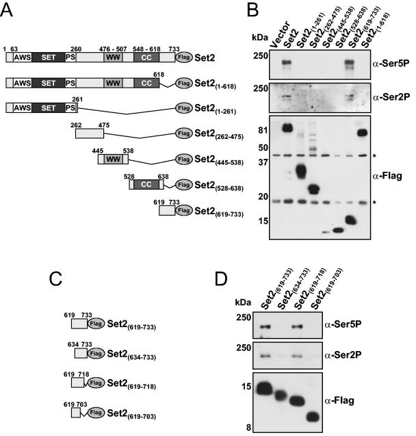
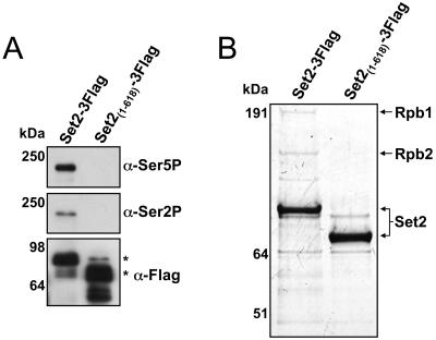
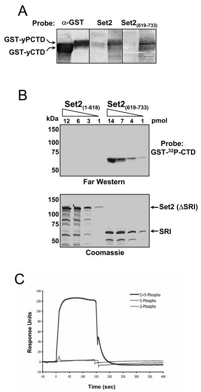
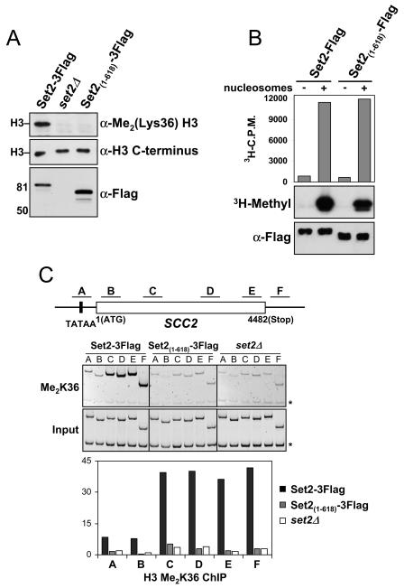
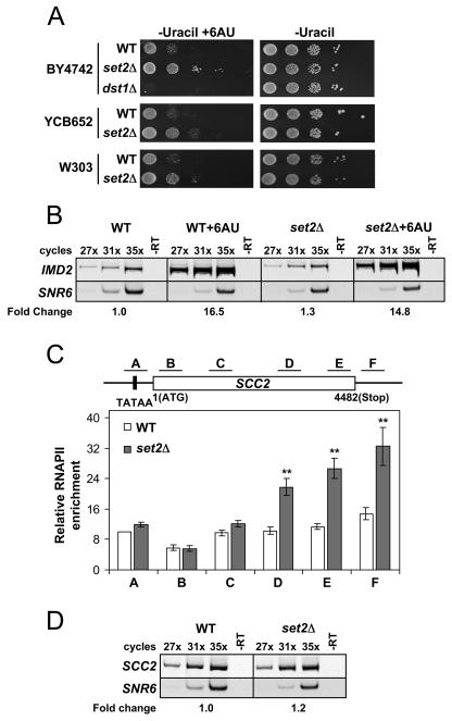
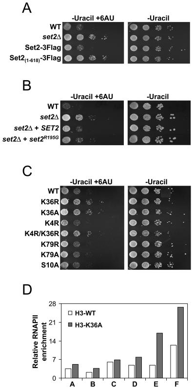
Histone methylation and the enzymes that mediate it are important regulators of chromatin structure and gene transcription. In particular, the histone H3 lysine 36 (K36) methyltransferase Set2 has recently been shown to associate with the phosphorylated C-terminal domain (CTD) of RNA polymerase II (RNAPII), implying that this enzyme has an important role in the transcription elongation process. Here we show that a novel domain in the C terminus of Set2 is responsible for interaction between Set2 and RNAPII. This domain, termed the Set2 Rpb1 interacting (SRI) domain, is encompassed by amino acid residues 619 to 718 in Set2 and is found to occur in a number of putative Set2 homologs from Schizosaccharomyces pombe to humans. Unexpectedly, BIACORE analysis reveals that the SRI domain binds specifically, and with high affinity, to CTD repeats that are doubly modified (serine 2 and serine 5 phosphorylated), indicating that Set2 association across the body of genes requires a specific pattern of phosphorylated RNAPII. Deletion of the SRI domain not only abolishes Set2-RNAPII interaction but also abolishes K36 methylation in vivo, indicating that this interaction is required for establishing K36 methylation on chromatin. Using 6-azauracil (6AU) as an indicator of transcription elongation defects, we found that deletion of the SRI domain conferred a strong resistance to this compound, which was identical to that observed with set2 deletion mutants. Furthermore, yeast strains carrying set2 alleles that are catalytically inactive or yeast strains bearing point mutations at K36 were also found to be resistant to 6AU. These data suggest that it is the methylation by Set2 that affects transcription elongation. In agreement with this, we have determined that deletion of SET2, its SRI domain, or amino acid substitutions at K36 result in an alteration of RNAPII occupancy levels over transcribing genes. Taken together, these data indicate K36 methylation, established by the SRI domain-mediated association of Set2 with RNAPII, plays an important role in the transcription elongation process.


Figures

FIG. 1.
FIG. 1.
Identification of a novel region in Set2 required for RNAPII binding. (A) Schematic representation of the Set2 constructs used to probe for RNAPII interaction. The SET domain along with the AWS domain, postSET domain (PS), WW domain, and coiled-coil motif (CC) are shown. All constructs contained a C-terminal Flag epitope. (B) set2Δ cells were transformed with either vector only or plasmids coding for the indicated Set2-Flag construct, and WCE were prepared. WCE were immunoprecipitated with anti-Flag beads followed by immunoblotting with antibodies directed against serine 5-phosphorylated CTD (H14; α-Ser5P), serine 2-phosphorylated CTD (H5; α-Ser2P), or the Flag epitope. Significant to mention is that the H5 antibody may also recognize serine 5 CTD phosphorylation in addition to serine 2 phosphorylation (16). Asterisks indicate the location of nonspecific Flag antibody-reactive species. (C) Schematic representation of the Set2-SRI domain constructs used to determine the boundaries of the functional SRI domain. N- and C-terminal truncations of the SRI domain were made in 15-amino-acid increments as shown. All constructs contained a C-terminal Flag epitope. (D) set2Δ cells were transformed with the indicated plasmids, WCE were prepared, and co-IPs were performed using the antibodies indicated in panel B. Sizes of the molecular mass markers are shown.
FIG. 2.
FIG. 2.
The SRI domain is required for interaction of Set2 with RNAPII. (A) Yeast strains containing full-length Set2 (Set2-3Flag) or a form of Set2 without the SRI domain [Set2(1-618)-3Flag] were made via genomic tagging with the 3xFlag epitope. WCEs of these strains were immunoprecipitated with anti-Flag beads followed by immunoblotting with antibodies directed against serine 2-phosphorylated CTD (H5; α-Ser2P), serine 5-phosphorylated CTD (H14; α-Ser5P), or the Flag epitope. Sizes of the molecular mass markers are shown, and asterisks indicate the location of expected Set2-Flag products. (B) WCEs from the strains in panel A were incubated with anti-Flag resin, and the resulting bound proteins were eluted with 3xFlag peptide. Eluted proteins were resolved by a 4-to-12% NuPAGE gel and examined by Coomassie staining. Arrows indicate the protein identity of bands in the Set2-3Flag lane that were examined by mass spectrometry, while analysis of parallel regions in the Set2(1-618)-3Flag lane was negative for the presence of Rpb1 or Rpb2. Sizes of the molecular mass markers are shown.
FIG. 3.
FIG. 3.
The SRI domain of Set2 binds synergistically to doubly modified CTD repeats. (A) Reverse far Western analysis. GST-yCTD and CTDK-I-phosphorylated GST-CTD (GST-yPCTD) fusion proteins were subjected to SDS-PAGE and transferred to nitrocellulose. Membranes were probed individually with purified recombinant full-length MBP-Set2 (Set2) or with MBP-SRI [Set2(619-733)], and the bound MBP fusions were detected with an anti-MBP antibody. As a control, a duplicate membrane was probed with an anti-GST antibody (α-GST) to demonstrate the presence of both GST-CTD fusion proteins. (B) Increasing amounts of two MBP fusion proteins [Set2(1-618) and Set2(619-733)] were resolved in two SDS-polyacrylamide gels; one gel was subjected to far Western analysis with GST-[32P]CTD as a probe, and the other was stained with Coomassie. (C) BIACORE analysis of the SRI domain. The MBP-SRI fusion protein [MBP-Set2(619-733)] was analyzed by surface plasmon resonance (BIACORE) for binding to distinct phosphorylated synthetic three-repeat CTD peptides. These peptides were either Ser5 phosphorylated (5-phospho), Ser2 phosphorylated (2-phospho), or both (2 + 5-phospho) in each repeat (see Materials and Methods). Response units, on the y axis, represent binding to the peptides. The binding response to a scrambled control peptide carrying six SerPO4residues (see Materials and Methods) has been subtracted from each of the three response curves. Only the peptide carrying both Ser2PO4 and Ser5PO4 in each repeat showed binding above control levels, and we estimate the affinity of this interaction (after subtraction of background binding to the control peptide) to be 6 μM.
FIG. 4.
FIG. 4.
Deletion of the SRI domain in Set2 abolishes H3 K36 dimethylation. (A) Yeast nuclear extracts prepared from set2Δ cells or the indicated genomically tagged strains in the BY4742 background were probed with antibodies against dimethylated lysine 36 at H3 [α-Me2(Lys36) H3] to monitor the role of the SRI domain in global K36 methylation levels. An antibody directed against the C terminus of H3 (α-H3 C terminus) was used as a loading control. Nuclear levels of Set2 in these strains were monitored using the anti-Flag antibody. A similar loss of K36 methylation was observed when the SRI domain was genomically deleted in the W303 background (data not shown). (B) Full-length (Set2-Flag) or SRI domain-truncated [Set2(1-618)-Flag] recombinant forms of Set2, which also contained an N-terminal MBP epitope, were prepared and analyzed for their HMT activity in vitro. HMT reactions were prepared with bacterial lysates containing the indicated Set2 constructs with or without nucleosomes. Identical samples were analyzed by the filter-binding assay (upper) and fluorography (middle). Immunoblotting with the Flag antibody (lower) was performed to ensure equal amounts of protein were present in each reaction mixture. (C) WT or SRI-deleted yeast strains were analyzed by ChIP for K36 dimethyl levels on genes. DNA isolated from the K36 methylation IPs was used in PCRs with primer pairs for the indicated regions of the SCC2 gene (top). PCR products of the input DNA (input) and ChIP DNA (Me2K36) are shown (middle). The asterisks indicate the location of a PCR product pertaining to an intergenic region at chromosome V (ChV), which was used as a loading control in all PCRs. The histogram displays the relative enrichment values for K36 dimethylation (bottom). The values were calculated by dividing the ratio of band intensities for IP DNA/ChV with the ratio of intensities for the input DNA/ChV. Identical results were found for the PMA1, ENO1, and ADH1 genes (data not shown).
FIG. 5.
FIG. 5.
Deletion of SET2 results in an elongation phenotype and an alteration of RNAPII occupancy on genes. (A) Various strains containing either WT, set2Δ, or dst1Δ alleles were plated on synthetic dextrose-uracil medium with or without 6AU (100 μg/ml) and grown at 30°C for 2 to 3 days to monitor for transcription elongation phenotypes. All strains contained the plasmid pRS316 containing the URA3 gene, except yeast strain YCB652, which contains an integrated URA3 gene. Results using mycophenolic acid (100 μg/ml) were found to yield identical results (data not shown). (B) The loss of Set2 does not aberrantly affect the increased expression of IMD2 in 6AU. Semiquantitative RT-PCR was used to monitor the expression of IMD2 and SNR6 (a polymerase III-transcribed gene used as a control) in WT or set2Δ strains in the absence or presence of 6AU (50 μg/ml). The results of RT-PCRs with (PCR amplification cycles indicated above) or without (-RT) reverse transcriptase are shown. The fold change in IMD2 expression under each condition is indicated, based on averages of the three cycling parameters for each strain, with WT set to 1.0 as reference. (C) WT or set2Δ strains were analyzed by ChIP for RNAPII levels. Isolated DNA from the RNAPII IPs were used in PCRs with primer pairs for regions of SCC2 as indicated in the schematic. The data shown represent the average of 13 individual ChIP assays from separate cell pellets. The standard error of the mean is indicated. Asterisks indicate the relative set2Δ RNAPII enrichment values that were statistically significant compared to their WT counterparts (P < 0.01 for primer set D and P < 0.001 for primer sets E and F). ChIP analyses using other RNAPII antibodies (8GW16 from Covance or Rpb3 from NeoClone) or the N terminus of Rpb1 (y-80; Santa Cruz) revealed the same pattern of RNAPII distribution as displayed in the figure (results not shown). (D) SCC2 expression was not changed in set2Δ despite the increase in RNAPII density detected by ChIP. The expression levels of SCC2 and SNR6 (control) were monitored by semiquantitative RT-PCR as described for panel B. The fold change of SCC2 expression in set2Δ cells compared to that in WT is displayed as described for panel C.
FIG. 6.
FIG. 6.
K36 methylation influences transcription elongation. (A) Genomically tagged strains containing either full-length Set2 (Set2-3Flag) or Set2 with the SRI domain deleted [Set2(1-618)-3Flag] were generated and assayed, as in Fig. 5A, for growth on 6AU and compared to WT and set2Δ strains. (B) set2Δ strains were transformed with a plasmid expressing WT Set2 (SET2) or a mutant form of Set2 which abolishes its catalytic activity (set2R195G) and assayed for growth on 6AU as before. (C) Yeast strains (WZY42 derived) bearing various point mutations on histone H3 were assayed for growth on 6AU as in Fig. 5A. (D) WT or a K36 mutated strain was analyzed by ChIP for RNAPII levels for the SCC2 gene, as in Fig. 5C. The histogram data are representative of two independent experiments which showed similar results.

References

    1. Ahn, S. H., M. Kim, and S. Buratowski. 2004. Phosphorylation of serine 2 within the RNA polymerase II C-terminal domain couples transcription and 3′ end processing. Mol. Cell 13:67-76. - PubMed
    1. Alen, C., N. A. Kent, H. S. Jones, J. O′Sullivan, A. Aranda, and N. J. Proudfoot. 2002. A role for chromatin remodeling in transcriptional termination by RNA polymerase II. Mol. Cell 10:1441-1452. - PubMed
    1. Archambault, J., F. Lacroute, A. Ruet, and J. D. Friesen. 1992. Genetic interaction between transcription elongation factor TFIIS and RNA polymerase II. Mol. Cell. Biol. 12:4142-4152. - PMC - PubMed
    1. Beisel, C., A. Imhof, J. Greene, E. Kremmer, and F. Sauer. 2002. Histone methylation by the Drosophila epigenetic transcriptional regulator Ash1. Nature 419:857-862. - PubMed
    1. Briggs, S. D., M. Bryk, B. D. Strahl, W. L. Cheung, J. K. Davie, S. Y. Dent, F. Winston, and C. D. Allis. 2001. Histone H3 lysine 4 methylation is mediated by Set1 and required for cell growth and rDNA silencing in Saccharomyces cerevisiae. Genes Dev. 15:3286-3295. - PMC - PubMed

Publication types

MeSH terms