Comparative Study
. 2005 Apr 25;92(8):1475-85.
doi: 10.1038/sj.bjc.6602495.

Ascites induces modulation of alpha6beta1 integrin and urokinase plasminogen activator receptor expression and associated functions in ovarian carcinoma

Affiliations
Comparative Study

Ascites induces modulation of alpha6beta1 integrin and urokinase plasminogen activator receptor expression and associated functions in ovarian carcinoma

N Ahmed et al. Br J Cancer. .

Abstract

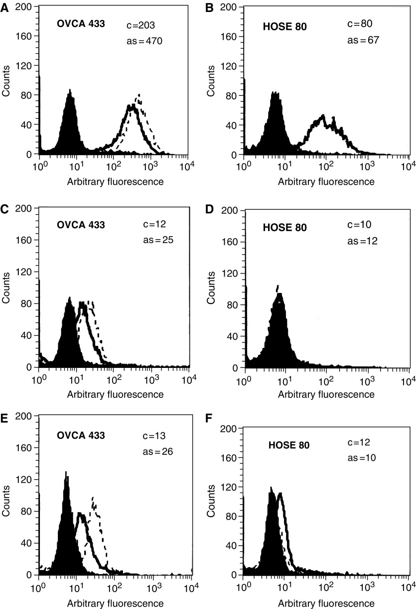
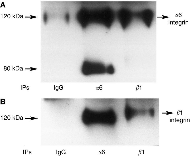
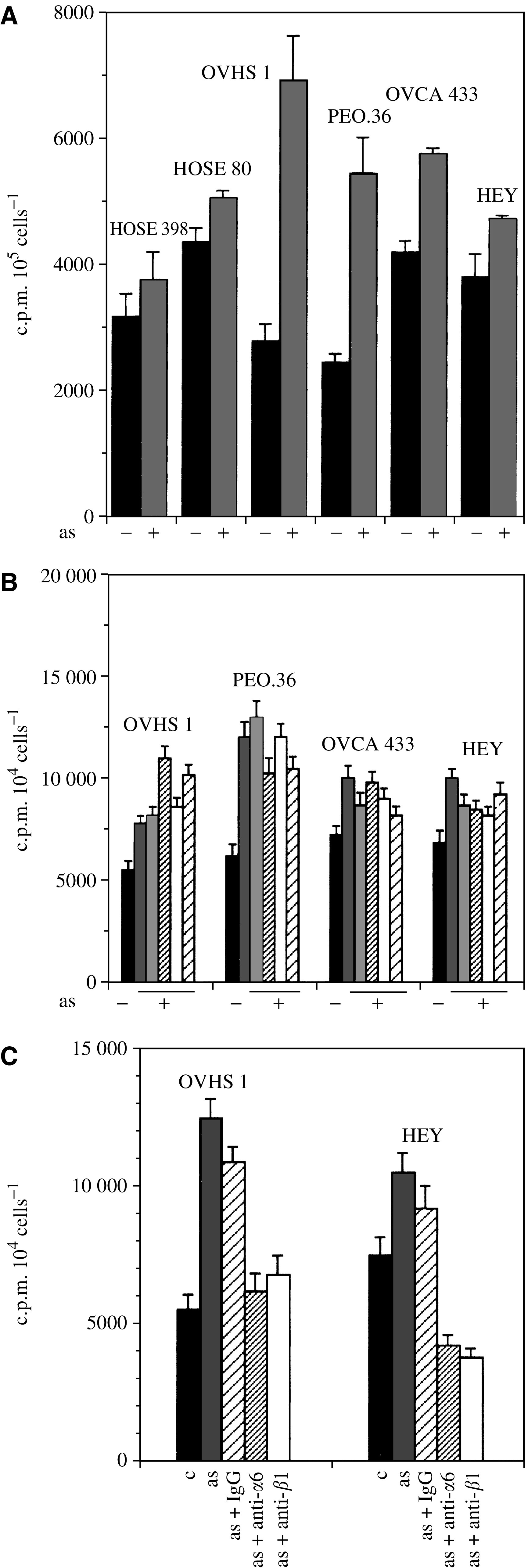
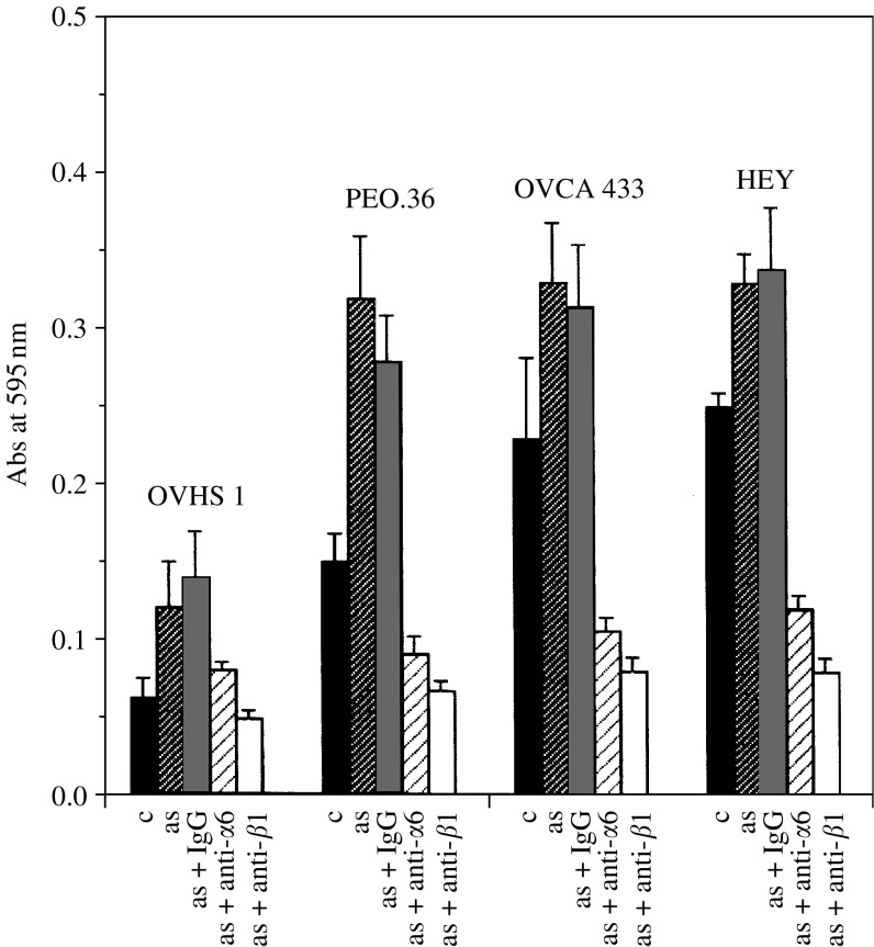
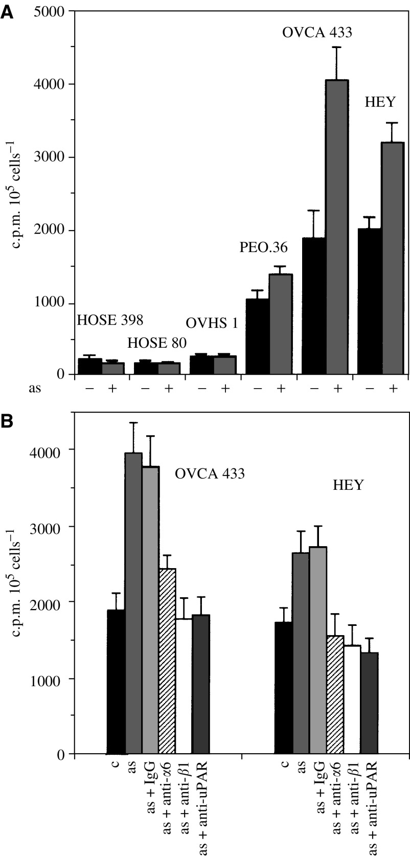
Interactions between cancer cells and the surrounding medium are not fully understood. In this study, we demonstrate that ascites induces selective changes in the expression of integrins and urokinase plasminogen activator/urokinase plasminogen activator receptor (uPA/uPAR) in ovarian cancer cells. We hypothesise that this change of integrin and uPA/uPAR expression triggers signalling pathways responsible for modulating phenotype-dependent functional changes in ovarian cancer cells. Human ovarian surface epithelial (HOSE) cell lines and epithelial ovarian cancer cell lines were treated with ascites for 48 h. Ascites induced upregulation of alpha6 integrin, without any change in the expression of alphav, beta1 and beta4 integrin subunits. Out of the four ovarian cancer cell lines studied, ascites induced enhancement in the expression of uPA/uPAR in the more invasive OVCA 433 and HEY cell lines without any change in the noninvasive OVHS1 and moderately invasive PEO.36 cell lines. On the other hand, no change in the expression of alpha6 integrin or uPAR, in response to ascites, was observed in HOSE cells. In response to ascites, enhancement in proliferation and in adhesion was observed in all four ovarian cancer cell lines studied. In contrast, no significant increase in proliferation or adhesion by ascites was observed in HOSE cells. Ascites-induced expression of uPA/uPAR correlated with the increased invasiveness of HEY and OVCA 433 cell lines but was not seen in OVHS1, PEO.36 and HOSE cell lines. Upregulation of alpha6 integrin and uPA/uPAR correlated with the activation of Ras and downstream Erk pathways. Ascites-induced activation of Ras and downstream Erk can be inhibited by using inhibitory antibodies against alpha6 and beta1 integrin and uPAR, consistent with the inhibition of proliferation, adhesion and invasive functions of ovarian cancer cell lines. Based on these findings, we conclude that ascites can induce selective upregulation of integrin and uPA/uPAR in ovarian cancer cells and these changes may modulate the functions of ovarian carcinomas.


Figures

Figure 1
Figure 1
Flow cytometric analyses of (A, B) α6 integrin; (C, D) uPAR and (E, F) uPA in OVCA 433 and HOSE 80 cell lines in response to ascites (3 mg ml−1). The median intensity of fluorescence was measured (MIF, arbitrary units, log scale) and is shown in the inserts of each cell line. The filled histogram in each figure is of control IgG, black lines indicate the expression of protein in the absence of ascites (C), while broken lines demonstrate protein expression in the presence of ascites (as) after 48 h. Results are representative of five experiments.
Figure 2
Figure 2
α6β1 interaction in HEY cells. Monolayer cultures of HEY cancer cell line was washed twice with PBS and harvested with trypsin-versene. Cells were lysed in lysis buffer (100 mM Tris-HCl, pH 7.5, 150 mM NaCl, 1 mM CaCl2, 1% Triton X-100, 0.1% SDS, 0.1% NP-40, 1 mM vanadate, 1 μg ml−1 pepstatin, 1 mM PMSF, 5 μg ml−1 Trasylol and I μg ml−1 of leupeptin). Protein concentrations of the cell lysates were determined and lysates containing equal protein were used for immunoprecipitation. Cells lysates were immunoprecipitated with mAbs against against α6 (43B-9B), β1 (PD52) or isotype matched control. Samples were resolved in 7.5% SDS–PAGE gel under nonreducing conditions and transferred to nitrocellulose membranes. Membranes were then probed with (A) anti-α6 integrin; (B) anti-β1 integrin antibodies and the interaction of α6β1 integrin was evaluated with ECL.
Figure 3
Figure 3
Expression of α6 integrin and uPAR in normal ovaries and ovarian tumours. Cryostat sections of ovarian tissues were stained by the immunoperoxidase method for the expression of α6 integrin and uPAR as discussed in the Materials and Methods section. (A) Normal ovary, arrow showing continuous basal expression of α6 integrin in the epithelium; (B) normal ovary, showing no expression of uPAR; (C) grade 3 serous ovarian tumour, arrows indicating irregular expression of α6 integrin in the basal epithelial and (D) grade 3 serous ovarian tumour, arrows indicating cytoplasmic epithelial staining of uPAR.
Figure 4
Figure 4
Effect of ascites on the proliferation of HOSE and ovarian cancer cells. The proliferative response of HOSE and ovarian cancer cells was determined on monolayer cultures and by 3D collagen gel system as described in Materials and Methods. (A) HOSE and ovarian cancer cell lines were grown as monolayers on 24-well plates and treated with ±ascites (3 mg ml−1) and the uptake of [3H]thymidine was measured over 16 h as described in Materials and Methods. The experiment was performed three times in triplicate. (B) Ovarian cancer cell lines grown in 3D collagen gel system were treated with five different ascites samples and their proliferation response was measured. The experiment was performed three times in triplicate. (C) Effect of α6 and β1 integrin inhibitory antibodies on the ascites-induced proliferation of OVHS1 and HEY cell lines in 3D collagen gel system.
Figure 5
Figure 5
Effect of ascites on the adhesion of HOSE and ovarian cancer cell lines. The adhesive response of ovarian cancer cells in response to ascites was determined as described in Materials and Methods. Ovarian cancer cell lines were treated for 24 h with ±ascites (3 mg ml−1), cells were washed and adhesion of the cells was measured using 96-well plates. The experiment was repeated three times in triplicate.
Figure 6
Figure 6
Effect of ascites on the invasion of HOSE and ovarian cancer cell lines. The invasive potential of HOSE and ovarian cancer cells was determined by Matrigel Boyden chamber as described in Materials and Methods. (A) HOSE and ovarian cancer cell lines were treated with ±ascites (3 mg ml−1) and their invasion capacity was measured in serum-free medium using 5% FBS as chemoattractant. The experiment was performed three times in triplicate. (B) Effect of α6, β1 integrin and uPAR inhibitory antibodies on the ascites-induced invasion of OVCA 433 and HEY cell lines.
Figure 7
Figure 7
Effect of ascites on the activation of Ras and Erk pathway in HEY and OVHS 1 cell lines. Activation of Ras and downstream Erk was performed as described in Materials and Methods. (A, D) HEY and OVHS 1 cells were serum starved for 16 h and then treated with ascites in the presence or absence of inhibitory antibodies for 30 min. Cells were lysed and Ras-GTP and Ras-GDP were immunoprecipitated as described in Materials and Methods. Equal amount of protein was loaded in each lane. Lanes 1, 3, 5, 7 and 9 demonstrate expression of Ras-GTP, while lanes 2, 4, 6, 8 and 10 demonstrate the expression of Ras-GDP. (B, E) Effect of ascites and α6, β1 integrin and uPAR inhibitory antibodies on the ascites-induced activation of Erk in HEY and OVHS 1 cells. (C, F) β-Tubulin staining of the cell lysate to show protein loading.

References

    1. Aguirre Ghiso JA, Kovalski K, Ossowski L (1999) Tumor dormancy induced by downregulation of urokinase receptor in human carcinoma involves integrin and MAPK signaling. J Cell Biol 147: 89–104 - PMC - PubMed
    1. Ahmed N, Niu J, Dorahy DJ, Gu X, Andrews S, Meldrum CJ, Scott RJ, Baker MS, Macreadie IG, Agrez MV (2002a) Direct integrin alphavbeta6-ERK binding: implications for tumour growth. Oncogene 21: 1370–1380 - PubMed
    1. Ahmed N, Oliva K, Wang Y, Quinn M, Rice G (2003a) Downregulation of urokinase plasminogen activator receptor expression inhibits Erk signalling with concomitant suppression of invasiveness due to loss of uPAR–beta1 integrin complex in colon cancer cells. Br J Cancer 89: 374–384 - PMC - PubMed
    1. Ahmed N, Pansino F, Clyde R, Murthi P, Quinn MA, Rice GE, Agrez MV, Mok S, Baker MS (2002b) Overexpression of alphavbeta6 integrin in serous epithelial ovarian cancer regulates extracellular matrix degradation via the plasminogen activation cascade. Carcinogenesis 23: 237–244 - PubMed
    1. Ahmed N, Riley C, Oliva K, Stutt E, Rice GE, Quinn MA (2003b) Integrin-linked kinase expression increases with ovarian tumour grade and is sustained by peritoneal tumour fluid. J Pathol 201: 229–237 - PubMed

Publication types

MeSH terms

Substances