Comparative Study
. 2005 Mar 30;25(13):3442-51.
doi: 10.1523/JNEUROSCI.0204-05.2005.

AMPA/kainate receptor-mediated downregulation of GABAergic synaptic transmission by calcineurin after seizures in the developing rat brain

Affiliations
Comparative Study

AMPA/kainate receptor-mediated downregulation of GABAergic synaptic transmission by calcineurin after seizures in the developing rat brain

Russell M Sanchez et al. J Neurosci. .

Abstract

Hypoxia is the most common cause of perinatal seizures and can be refractory to conventional anticonvulsant drugs, suggesting an age-specific form of epileptogenesis. A model of hypoxia-induced seizures in immature rats reveals that seizures result in immediate activation of the phosphatase calcineurin (CaN) in area CA1 of hippocampus. After seizures, CA1 pyramidal neurons exhibit a downregulation of GABA(A) receptor (GABA(A)R)-mediated inhibition that was reversed by CaN inhibitors. CaN activation appears to be dependent on seizure-induced activation of Ca2+-permeable AMPA receptors (AMPARs), because the upregulation of CaN activation and GABA(A)R inhibition were attenuated by GYKI 52466 [1-(4-aminophenyl)-4-methyl-7,8-methylenedioxy-5H-2,3-benzodiazepine hydrochloride] or Joro spider toxin. GABA(A)R beta2/3 subunit protein was dephosphorylated at 1 h after seizures, suggesting this subunit as a possible substrate of CaN in this model. Finally, in vivo administration of the CaN inhibitor FK-506 significantly suppressed hypoxic seizures, and posttreatment with NBQX (2,3-dihydroxy-6-nitro-7-sulfonyl-benzo[f]quinoxaline) or FK-506 blocked the hypoxic seizure-induced increase in CaN expression. These data suggest that Ca2+-permeable AMPARs and CaN regulate inhibitory synaptic transmission in a novel plasticity pathway that may play a role in epileptogenesis in the immature brain.


Figures

Figure 1.
Figure 1.
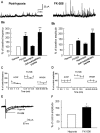
GABAAR-mediated sIPSCs and eIPSCs were significantly deceased in CA1 pyramidal neurons after hypoxia-induced seizures. A, Representative traces show outward sIPSCs with cells voltage clamped at 10 mV. The sIPSC frequency and amplitudes clearly were decreased in slices removed at 1 h after hypoxia-induced seizures in P10 rat pups compared with slices from littermate controls. The addition of bicuculline methiodide completely abolished spontaneous events, confirming that the synaptic currents were mediated by GABAARs. B, Pooled data (mean ± SEM) showed a significant decrease in sIPSC frequency and amplitude in slices from hypoxia-treated pups (0.82 ± 0.1 Hz, **p < 0.01; 12.9 ± 1.4 pA; n = 5 cells per group; *p < 0.05) compared with controls (1.68 ± 0.2 Hz and 19.1 ± 1.5 pA; n = 5; Student's t test). (Data shown are normalized to control values.) Ca, Superimposed representative traces show that eIPSCs induced by single stimulus shocks were markedly reduced after hypoxia-induced seizures. Cb, The amplitudes of eIPSCs were significantly decreased in slices from hypoxia-treated rats (13.7 ± 1.4 pA; n = 9) compared with control rats (45.1 ± 4.7 pA; n = 8; ***p < 0.0001). D, Current-voltage plots for GABA-evoked currents showed no difference in the reversal potential between the control group (Da) and hypoxia-treated group (Db), indicating that a persistent shift in reversal potential could not have explained the difference in IPSC amplitudes between groups. Traces in the inset are current responses to 10 s applications of GABA (100 μm) recorded under the same conditions as eIPSCs.
Figure 2.
Figure 2.
A, Inhibition of CaN activity reversed the decreases in sIPSC amplitude and frequency in hippocampal CA1 pyramidal neurons after hypoxia-induced seizures. B, sIPSC frequency (0.82 ± 0.1 Hz) and amplitudes (12.9 ± 1.4 pA) in cells from the hypoxia-treated group were significantly increased by application of FK-506, 1.77 ± 0.19 Hz (**p < 0.01) and 17.3 ± 0.8 pA (*p < 0.05; before drug, n = 5; FK-506, n = 4), respectively. sIPSC frequency (0.85 ± 0.06 Hz) and amplitudes (7.7 ± 0.6 pA) also were significantly increased after CaN autoinhibitory peptide application to 2.2 ± 0.28 Hz and 15.8 ± 1.5 pA, respectively (n = 6; ***p < 0.001). In contrast, no significant changes were observed in sIPSC frequency and amplitudes after rapamycin application (prerapamycin values were 0.88 ± 0.08 Hz and 9.5 ± 0.7 pA, n = 6, and postrapamycin values were 0.93 ± 0.08 Hz and 10.2 ± 0.6 pA, n = 6, for frequency and amplitude, respectively). C, D, The effects of bath application of FK-506 recovered with washout. E, Superimposed representative traces show that the decreased eIPSC amplitude after hypoxia-induced seizures also was significantly reversed by bath application of FK-506. F, Pooled data showed that FK-506 treatment significantly reversed the eIPSC amplitude (n = 6; *p < 0.05). The mean amplitudes of eIPSCs were 13.7 ± 1.4 pA (n = 9) before FK-506 and 20.8 ± 1.3 pA (n = 6) after FK-506.
Figure 3.
Figure 3.
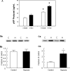
Increased CaN phosphatase activity after hypoxia-induced seizures. A, Hypoxia-induced seizures resulted in a significant increase in control basal CaN (c basal) and maximal control CaN (c max) activity. Basal and maximal CaN activity levels were assayed in homogenates from area CA1 of hippocampus at 1 h after hypoxia. CaN activity was significantly increased in hypoxia samples (black, h basal, h max) compared with control samples (white, c basal, c max) at both basal levels (*p < 0.001; n = 6; Student's t test) and Mn2+ stimulated maximal levels (*p < 0.001; n = 6; Student's t test). Western blot analyses of CaN protein expression were performed in hippocampal homogenates reacted with monoclonal CaN-Aα antibody at 1 h (Ba) and 24 h (Ca) after hypoxia-induced seizures (H) and compared with controls (C). Bb, Histogram from pooled data shows that at 1 h after hypoxia, hippocampal CaN protein expression was not significantly different from that of controls (n = 5; p > 0.05), whereas hippocampal homogenates removed at 24 h (Cb) after hypoxia-induced seizures showed a significant increase in CaN protein compared with controls (n = 6; *p < 0.05).
Figure 4.
Figure 4.
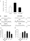
A, GYKI 52466 prevented the hypoxia-induced increase in CaN activity. CaN activity was significantly increased after hypoxia-induced seizures (n = 14; *p < 0.05), whereas pre-incubation of hippocampal homogenates with GYKI 52466 (30 μm) after hypoxia in vivo prevented this increase. GYKI 52466 caused no significant difference in CaN activity from the control samples (n = 5). B, C, Blockade of Ca2+-permeable AMPARs reversed sIPSC downregulation, where as blockade of NMDARs did not affect sIPSCs after hypoxia-induced seizures. Application of GYKI 52466 increased sIPSC frequency and amplitude from 0.76 ± 0.03 Hz and 7.5 ± 0.7 pA (hypoxia group) to 1.42 ± 0.15 Hz (n = 5; p < 0.001) and 10.1 ± 0.7 pA (before drug, n = 6; after GYKI 52466, n = 5; p < 0.05), respectively, whereas JSTx increased the frequency from 0.80 ± 0.07 Hz to 1.44 ± 0.19 Hz (n = 6; p < 0.01) and amplitude from 7.1 ± 0.7 to 11.1 ± 1.0 pA (n = 6; p < 0.01). d-AP-5 did not significantly affect the decreased sIPSC frequency [frequencies were 0.85 ± 0.05 Hz before drug and 0.94 ± 0.05 Hz after drug (n = 5); amplitudes were 9.3 ± 0.8 pA before drug and 10.1 ± 1.1 pA after drug (n = 5; p > 0.05)].
Figure 5.
Figure 5.
Regulation of sEPSCs in CA1 pyramidal neurons after hypoxia-induced seizures at P10. A, Representative traces show that the sEPSC frequency (but not amplitude) was significantly increased after hypoxia-induced seizures. B, Summary of the regulation of sEPSCs after hypoxic seizures from the pooled data shows that the sEPSC frequency was significantly increased from 1.13 ± 0.17 Hz (n = 4) to 1.94 ± 0.19 Hz (n = 7; *p < 0.05), whereas the amplitude was not significantly changed, with values of 24.3 ± 9.2 pA (n = 4) for controls and 18.8 ± 4.4 pA (n = 7) for the hypoxia-treated group.
Figure 6.
Figure 6.
Effect of FK-506 treatment on seizure incidence during perinatal hypoxia. Rat pups received doses of FK-506 (10 mg/kg, i.p.). A, Pretreatment of FK-506 30 min before exposure to global hypoxia; FK-506 significantly decreased the seizure incidence (n = 12) induced by hypoxia compared with control vehicle injection (hypoxia alone) (n = 11; **p < 0.001). B, Immediately after hypoxia, animals received injections of FK-506 or saline vehicle. Control animals also received injections of saline vehicle at this time. Injections were repeated at 8 and 16 h after hypoxia. The total number of seizures was significantly increased in the hypoxic group (n = 10) compared with control animals (n = 11), and FK-506 significantly blocked this effect (n = 10; *p < 0.05). C, FK-506 treatment (n = 8) blocked the significant increases in cumulative seizure duration (n = 7) compared with controls (n = 6; *p < 0.005). D, In addition, the average length of a seizure was also significantly increased in the hypoxic group (n = 7) for animals that seized during hypoxia at P11 compared with controls (n = 6) and FK-506-treated animals (n = 8; **p < 0.05).
Figure 7.
Figure 7.
CaN expression was significantly increased 24 h for the hypoxia group (H) (156.2 ± 12.8% of control; n = 8; p < 0.05). FK-506 treatment (F) (10 mg/kg) 30 min before hypoxia and 6 h after hypoxia reversed CaN expression to control (C) levels (98.6 ± 9.7% of control; n = 5; p < 0.05). NBQX treatment (N) (20 mg/kg) immediately after and at 12 h after hypoxia also suppressed the increase in CaN expression (94.9 ± 8.1% of the control; n = 4; p < 0.05) compared with the saline-treated hypoxic pups.

Comment in

Similar articles

Cited by

References

    1. Bergamasco B, Penna P, Ferrero P, Gavinelli R (1984) Neonatal hypoxia and epileptic risk: a clinical prospective study. Epilepsia 25: 131-146. - PubMed
    1. Bernes SM, Kaplan AM (1994) Evolution of neonatal seizures. Pediatr Clin North Am 41: 1069-1104. - PubMed
    1. Blair RE, Churn SB, Sombati S, Lou JK, DeLorenzo RJ (1999) Long-lasting decrease in neuronal Ca2+/calmodulin-dependent protein kinase II activity in a hippocampal neuronal culture model of spontaneous recurrent seizures. Brain Res 851: 54-65. - PubMed
    1. Brandon NJ, Uren JM, Kittler JT, Wang H, Olsen R, Parker PJ, Moss SJ (1999) Subunit-specific association of protein kinase C and the receptor for activated C kinase with GABA type A receptors. J Neurosci 19: 9228-9234. - PMC - PubMed
    1. Brandon NJ, Delmas P, Kittler JT, McDonald BJ, Sieghart W, Brown DA, Smart TG, Moss SJ (2000) GABAA receptor phosphorylation and functional modulation in cortical neurons by a protein kinase C-dependent pathway. J Biol Chem 275: 38856-38862. - PubMed

Publication types

MeSH terms

LinkOut - more resources